A multiplex fluorescent gene detection method for rapidly distinguishing ect, mad, mcmv, poly

A multiple fluorescence, rapid technology, applied in the direction of microbial determination/inspection, biochemical equipment and methods, etc., to achieve the effect of reducing detection cost, less sample consumption, and simple operation

Active Publication Date: 2018-07-24
GUANGDONG LAB ANIMALS MONITORING INST
View PDF3 Cites 0 Cited by
  • Summary
  • Abstract
  • Description
  • Claims
  • Application Information

AI Technical Summary

Problems solved by technology

Compared with conventional PCR, fluorescent quantitative PCR has advantages in sensitivity, specificity, and speed. However, real-time fluorescent PCR technology is limited by the type of fluorescence and the instrument itself, and can only detect up to 5 targets.

Method used


Image

  • A multiplex fluorescent gene detection method for rapidly distinguishing ect, mad, mcmv, poly
  • A multiplex fluorescent gene detection method for rapidly distinguishing ect, mad, mcmv, poly
  • A multiplex fluorescent gene detection method for rapidly distinguishing ect, mad, mcmv, poly

Examples


example 1

[0035] Construction of example 1 murine pox virus, mouse adenovirus, mouse cytomegalovirus and mouse polyomavirus plasmid

[0036] Use the kit TIANamp Genomic DNA Kit to extract the DNA of ECT, MAD, MCMV and POLY viruses respectively, and perform PCR amplification with the above corresponding primers respectively. The amplification primers are:

[0037] ECT primer E1: 5'-TACGGTGTATATGGCTACTCA-3' (SEQ ID NO: 1);

[0038] ECT primer E2: 5'-ACTGCTGCTTCCTATACTCG-3' (SEQ ID NO: 2);

[0039] MAD primer A1: 5'-AGACTCACACTGCGTATAGTCC-3' (SEQ ID NO: 3);

[0040] MAD primer A2: 5'-CCAGAACTCGGTTATTCCCCA-3' (SEQ ID NO: 4);

[0041] MCMV Primer C1: 5'-GCCCTCTTCATCCCTTACCT-3' (SEQ ID NO: 5);

[0042] MCMV primer C2: 5'-GACCTCCATGTCCTTCATGC-3' (SEQ ID NO: 6).

[0043] POLY primer P1: 5'-CCATGCTCCCTTCTATGCGAT-3' (SEQ ID NO: 7);

[0044] POLY primer P2: 5'-TCAGTCAGCCATAGATGCAA-3' (SEQ ID NO: 8).

[0045] The 5' ends of primers E1, A1, C1 and P1 are also added with a tag sequence through th...

Embodiment 2

[0069] Example 2: Establishment of multiple fluorescent gene detection methods for mousepox virus, mouse adenovirus, mouse cytomegalovirus and mouse polyomavirus:

[0070] 1) Plasmid PCR amplification

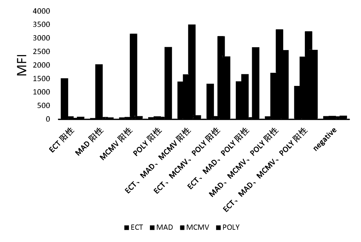
[0071] Singleplex, triplex, and quadruple PCR amplification with primers that specifically amplify mousepox virus, mouse adenovirus, mouse cytomegalovirus, and mouse polyomavirus.

[0072] Preparation of upstream primer mixture: mix E1, A1, C1 and P1 at a ratio of 1:1:1:1; preparation of downstream primer mixture: mix E2, A2, C2 and P2 at a ratio of 1:1:1:1 ratio to mix. The specific regions of the above four viruses were amplified by using four specific templates, triple templates, and quadruple templates. The preparation of the triple template: mixing the three plasmids in a ratio of 1:1:1; the preparation of the quadruple template: mixing the four plasmids in a ratio of 1:1:1:1. The PCR amplification system and procedure were the same as in Example 1.

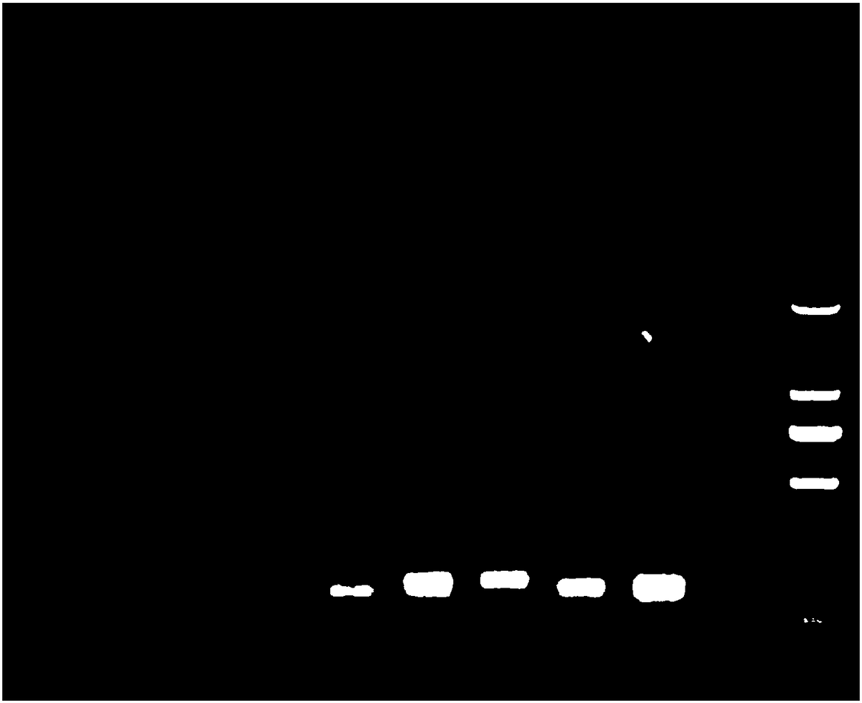
[0073] PCR produc...

Embodiment 3

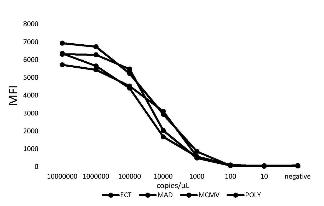
[0084] Embodiment 3: Multiple fluorescent gene detection sensitivity experiment of ECT, MAD, MCMV, POLY

[0085] The prepared plasmid was quantified, diluted by 10-fold dilution method to 10 copies / μL, and detected by the multiple fluorescent gene detection method established above. The sensitivity test results of multiple fluorescent gene detection of ECT, MAD, MCMV and POLY are as follows: image 3 As shown, the experimental results show that the sensitivity of ECT, MAD, MCMV, and POLY is high, and the detection limit is 1000copies / μL.


PUM


Abstract

The invention discloses a multiplex fluorescence gene detection method for quickly distinguishing ECT, MAD, MCMV and POLY. The method is simple in operation and a target amplification fragment is obtained through PCR. After that, an amplification product, fluorescent encoding microspheres and streptavidin-phycoerythrin are subjected to hybridization, and then an MFI value is read by a detector. In this way, different types of viruses can be distinguished. According to the technical scheme of the invention, the method is good in sensitivity, accuracy, repeatability and specificity, and simple, convenient and quick to use. Compared with the conventional detection methods, the above method realizes the simultaneous detection of different target fragments in the same sample. Meanwhile, the method has the advantages of small sample amount, and simple and rapid operation. The detection cost can be greatly reduced.

Description

technical field [0001] The invention belongs to the field of virus detection of experimental animals, in particular to a multiple fluorescent gene detection method for rapidly distinguishing ECT, MAD, MCMV and POLY. Background technique [0002] Mousepox virus (Mousepox virus), also known as Ectromelia virus (ECT), can cause severe infection in experimental mice. It is divided into acute and chronic types, and most of them are recessive infections. The disease is often explosive and has a high fatality rate, often resulting in the elimination of the whole group, which brings great harm to scientific research and production. It is a must-test item for clean-grade mice. Murine Adenovirus (MAD) is a new type of animal virus with double-stranded DNA isolated from mice in the 1960s. It is widely distributed and can easily cause respiratory tract infections, such as acute febrile pharyngitis. , pharyngoconjunctival fever and pneumonia, etc., are pathogens that need to be excluded...

Claims


Application Information

Patent Timeline
Patent Type & Authority Patents(China)
IPC IPC(8): C12Q1/70C12Q1/686
CPCC12Q1/6816C12Q1/701C12Q2537/143C12Q2563/149C12Q2563/107
Inventor 尹雪琴郭鹏举黄韧
Owner GUANGDONG LAB ANIMALS MONITORING INST
Why Eureka
Try Eureka