Three-dimensional imaging device for detecting human body microvascular ultramicrostructure

An ultrastructure and three-dimensional imaging technology, applied in the field of medical devices, can solve the problems that two-dimensional imaging cannot meet the requirements, and two-dimensional imaging cannot obtain depth information, etc., and achieve the effect of improving clarity, simple and compact structure, and convenient operation

Pending Publication Date: 2017-04-26
QINGDAO UNIV
View PDF10 Cites 2 Cited by
  • Summary
  • Abstract
  • Description
  • Claims
  • Application Information

AI Technical Summary

Problems solved by technology

Although the analysis of two-dimensional imaging can quantify and digitize the flow velocity of microvessels, the diameter of microvessels, and the density of microvessels in cross-section, but because two-dimensional imaging cannot obtain depth information, the microvessel density and shape of microvessels in longitudinal section analysis, 2D imaging does not meet the requirements

Method used


Image

  • Three-dimensional imaging device for detecting human body microvascular ultramicrostructure
  • Three-dimensional imaging device for detecting human body microvascular ultramicrostructure
  • Three-dimensional imaging device for detecting human body microvascular ultramicrostructure

Examples


Embodiment 1

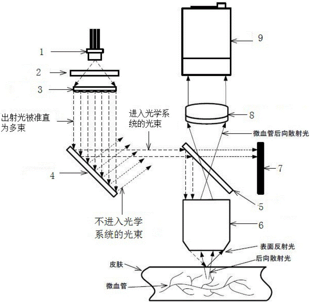
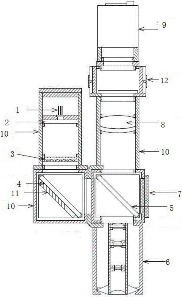
[0051] Such as Figure 2-3A three-dimensional imaging device for detecting the ultrastructure of human microvessels shown at least includes: a light source module for emitting multiple beams of collimated and parallel outgoing light; a light adjustment unit 2 for adjusting the outgoing light to circularly polarized light; The array reflective unit 4 for sequentially irradiating multiple beams of outgoing light into the dark field objective lens 6; the spectroscopic unit 5 for partially reflecting the light and partially transmitting it; Dark field objective lens 6; imaging unit 8 for imaging; imaging module 9 for presenting images.

[0052] Further, the imaging unit is used to focus light into the lens unit and receive the imaging light of the lens unit to make it clearly imaged on the imaging unit. The positions of the convex lenses can be interchanged, for example, a concave-convex lens and a biconvex lens are arranged in sequence along the direction of light source irradia...

Embodiment 2

[0072] Different from Example 1, as Figure 4-5 As shown, the dimming unit is arranged between the array transmission unit and the array reflection unit, that is, the outgoing light of the light source is collimated and divided into multiple beams, and then the beam is modulated into circularly polarized light. All the other are with embodiment 1.

[0073] In the method of using the three-dimensional imaging device to detect the microcirculation of the human body, the difference from Embodiment 1 is that the outgoing light of the light source is collimated and divided into multiple beams, and then the beam is modulated into circularly polarized light before entering the optical system. All the other are with embodiment 1.

Embodiment 3

[0075] Different from Example 1, as Figure 6-7 As shown, the dimming unit is arranged between the array reflection unit and the spectroscopic unit, that is, the adjusted light beam that needs to enter the optical system is modulated into circularly polarized light before entering the optical system. All the other are with embodiment 1.

[0076] In the method of using the three-dimensional imaging device to detect the microcirculation of the human body, the difference from Embodiment 1 is that the adjusted light beam that needs to enter the optical system is modulated into circularly polarized light before entering the optical system. All the other are with embodiment 1.


PUM


Abstract

The invention relates to a three-dimensional imaging device for detecting a human body microvascular ultramicrostructure. The device at least comprises a light source module used for transmitting multiple collimation and parallel emergent lights, a dimming unit used for adjusting the emergent light into circularly polarized light, an array reflection unit used for illuminating multiple emergent lights into a dark-field objective lens according to sequence, a light splitting unit used for partially reflecting and partially transmitting the light, the dark-field objective lens used for intensively illuminating an annular incident light to a focus position, an imaging unit used for imaging, and an image presenting module for presenting images. According to the three-dimensional imaging device, by matching of the array transmission unit, the array reflection unit and the dark-field objective lens, multiple emergent lights split from one light beam sequentially enter the dark-field objective lens, the dark-field objective lens is illuminated by incident lights at different angles on the same position and respectively imaged, and a three-dimensional image of microvessel is obtained by using a photometric three-dimensional measurement method, so that the microvessel density and microvessel shape of a longitudinal section are accurately subjected to digital quantification.

Description

technical field [0001] The invention relates to a medical device for medical diagnosis using optical imaging technology, in particular to a three-dimensional imaging device for detecting the ultrastructure of human microvessels. Background technique [0002] Microcirculation refers to the place where blood and tissue cells exchange substances between arterioles and venules. The integrity of the function, shape and metabolism of the microcirculation is an indispensable condition for maintaining the normal function of human organs. Through the study of microcirculation, it is convenient to further understand the special functions of various organs of the human body, and to recognize the pathogenesis of diseases, which is beneficial to disease prevention, diagnosis and treatment. A variety of disease states, including diabetes, hypertension and coronary heart disease, will cause pathological changes in microcirculation, including changes in microvascular diameter, microvascula...

Claims


Application Information

Patent Timeline
Patent Type & Authority Applications(China)
IPC IPC(8): A61B5/00
CPCA61B5/0059A61B5/489
Inventor 董蒨于綦悦魏宾夏楠
Owner QINGDAO UNIV
Why Eureka
Try Eureka