Imaging method of hard X-ray grating interferometer with single exposure

A technology of grating interferometry and imaging method, applied in the field of hard X-ray imaging physics, can solve the problems that hinder the popularization and application of hard X-ray grating interferometer, increase radiation dose and radiation damage risk, and long data acquisition time, etc., so as to reduce radiation damage risk, avoidance of high radiation doses, effects of reduced experiment time

Active Publication Date: 2017-05-10
HEFEI UNIV OF TECH
View PDF9 Cites 6 Cited by
  • Summary
  • Abstract
  • Description
  • Claims
  • Application Information

AI Technical Summary

Problems solved by technology

This method requires a complex horizontal step-scanning grating, which leads to a long data acquisition time and reduces the experimental efficiency; it requires the collection of multiple object projection images (not less than 4 in th

Method used


Image

  • Imaging method of hard X-ray grating interferometer with single exposure
  • Imaging method of hard X-ray grating interferometer with single exposure
  • Imaging method of hard X-ray grating interferometer with single exposure

Examples


Embodiment Construction

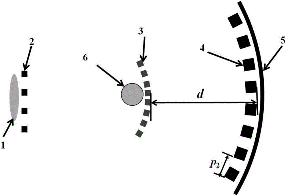
[0038] like figure 1 As shown, the hard X-ray grating interferometer includes: X-ray source 1, source grating 2, phase grating 3, analysis grating 4, detector 5; The imaging object 6; the source grating 2 is placed close to the X-ray source 1; the imaged object 6 is placed close to the inner side of the phase grating 3; an analysis grating 4 is arranged outside the phase grating 3; The axial distance is d; the detector 5 is closely attached to the outside of the analysis grating 4; in this embodiment, the imaging method of the hard X-ray grating interferometer for one exposure is carried out according to the following steps:

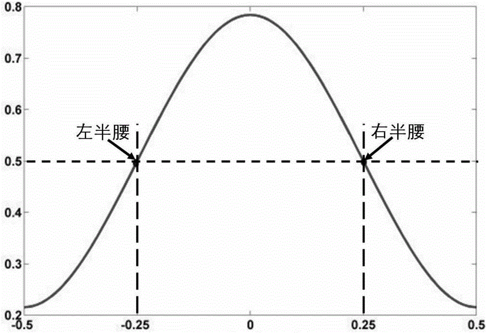
[0039] Step 1. Fix any two gratings in the source grating 2, the phase grating 3 and the analysis grating 4, and move the third grating along the moving direction by a quarter of the grating period, so that the hard X-ray grating interferometer works The point is fixed at the left half waist or right half waist position of the light intensity curve ( ...


PUM


Abstract

The invention discloses an imaging method of a hard X-ray grating interferometer with single exposure. The imaging method is characterized by including the steps of 1, moving any grating, and fixing a working point of the hard X-ray grating interferometer at a left half waist and right half waist position of a light intensity curve; 2, obtaining a background projection image and a projection image of an imaged object respectively; 3, obtaining the normalized projection image of the imaged object, and conducting logarithm process on the normalized projection image; 4, constructing an expression formula to conduct one-dimensional Fourier transform according to the logarithm process result; 5, utilizing one-dimensional inverse Fourier transform to extract absorption of the imaged object and a relative phase shift signal respectively according to the result of the Fourier transform. According to the imaging method of the hard X-ray grating interferometer with the single exposure, the data collection procedure of the hard X-ray grating interferometer is simplified, the imaged object is subjected to single exposure, the imaging efficiency is improved, the radiation damage risk is reduced, and thus a new path of the development of a future fast and low-radiation-dose clinical medical imaging technology is provided.

Description

technical field [0001] The invention relates to the field of hard X-ray imaging physics and methods, in particular to an imaging method of a hard X-ray grating interferometer with one exposure. Background technique [0002] Over the past 100 years, X-ray imaging methods and technologies have continuously made new progress in imaging physical mechanisms, data acquisition schemes, tomographic reconstruction algorithms, etc., and have now developed into the fields of materials science, clinical medicine, archaeology, and public safety. An indispensable tool. Especially in the field of clinical medicine, X-ray imaging methods and devices, including X-ray projection imaging (X-ray Radiography) and X-ray Computed Tomography (X-ray Computed Tomography), have made significant contributions to modern human health and social development. irreplaceable contribution. [0003] In terms of imaging physical mechanism, existing X-ray imaging methods and technologies are based on the diffe...

Claims


Application Information

Patent Timeline
IPC IPC(8): A61B6/00
Inventor 王志立刘达林
Owner HEFEI UNIV OF TECH
Why Eureka
Try Eureka