Marker for diagnosis of inflammation-associated HCC and application thereof

A technique for diagnosing markers and liver cancer, which is applied in the field of disease diagnosis and can solve problems such as unsatisfactory differentiation techniques

Active Publication Date: 2017-05-24
CENT FOR EXCELLENCE IN MOLECULAR CELL SCI CHINESE ACAD OF SCI
View PDF0 Cites 8 Cited by
  • Summary
  • Abstract
  • Description
  • Claims
  • Application Information

AI Technical Summary

Problems solved by technology

According to clinical practice, HCC is divided into two types: non-inflammation-related liver cancer and inflammation-related liver cancer. Different methods of clinical treatment should be adopted for these two types of liver cancer, but the technology for distinguishing these two types of liver cancer in the prior art is not ideal enough

Method used


Image

  • Marker for diagnosis of inflammation-associated HCC and application thereof
  • Marker for diagnosis of inflammation-associated HCC and application thereof
  • Marker for diagnosis of inflammation-associated HCC and application thereof

Examples


Embodiment 1

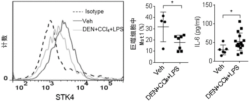
[0061] Example 1, STK4 protein and IL-6 protein levels in liver cancer mice

[0062] Using DEN+CCl 4 + LPS induces the process of inflammation-related liver cancer. DEN was injected intraperitoneally into 6-week-old wild-type mice. In the third week after injection, low-dose LPS was injected every day, and in the fourth week, CCl was given once a week. 4 Injection and observation after 12 weeks. After 11 months, the liver was removed, the lymphocytes in the liver were separated by Percoll, and the F4 / 80 and STK4 were stained by FACS flow cytometry, and the protein level of STK4 in macrophages was detected. At the same time, the blood was coagulated at room temperature for 60 minutes after eyeball blood collection, centrifuged at 3000 rpm / min for 20 minutes, and the supernatant serum was collected. The content of IL-6 in serum was detected by ELISA kit.

[0063] The result is as figure 1 , untreated normal control group (Veh) and DEN+CCl 4 There were significant difference...

Embodiment 2

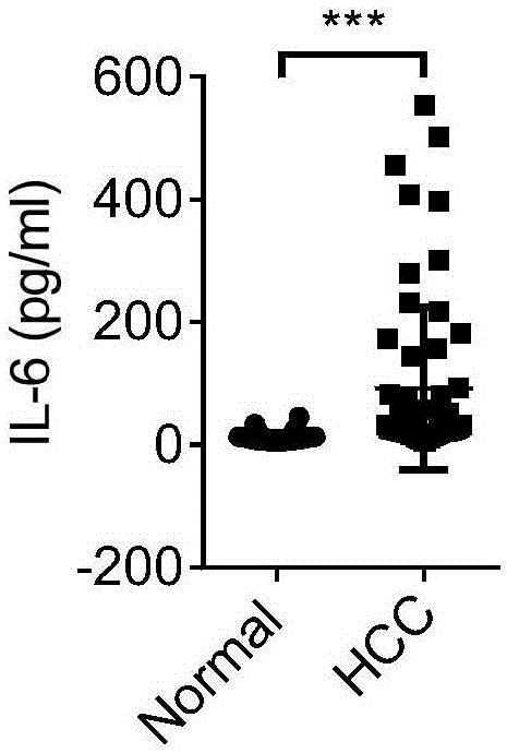
[0065] Example 2, the level of IL-6 in the peripheral blood of patients with liver cancer

[0066] Plasma was collected from 40 normal healthy people and 59 liver cancer patients. 3ml of blood from normal healthy people or patients with liver cancer collected in anticoagulant tubes was separated by Ficoll, and the uppermost layer of plasma was taken, and the content of IL-6 in the plasma was detected by ELISA.

[0067] The result is as figure 2 , The plasma level of IL-6 in patients with liver cancer was detected by ELISA method; while the level in normal healthy people was extremely low, the highest level was less than 20pg / ml, and there was a significant difference between the two.

Embodiment 3

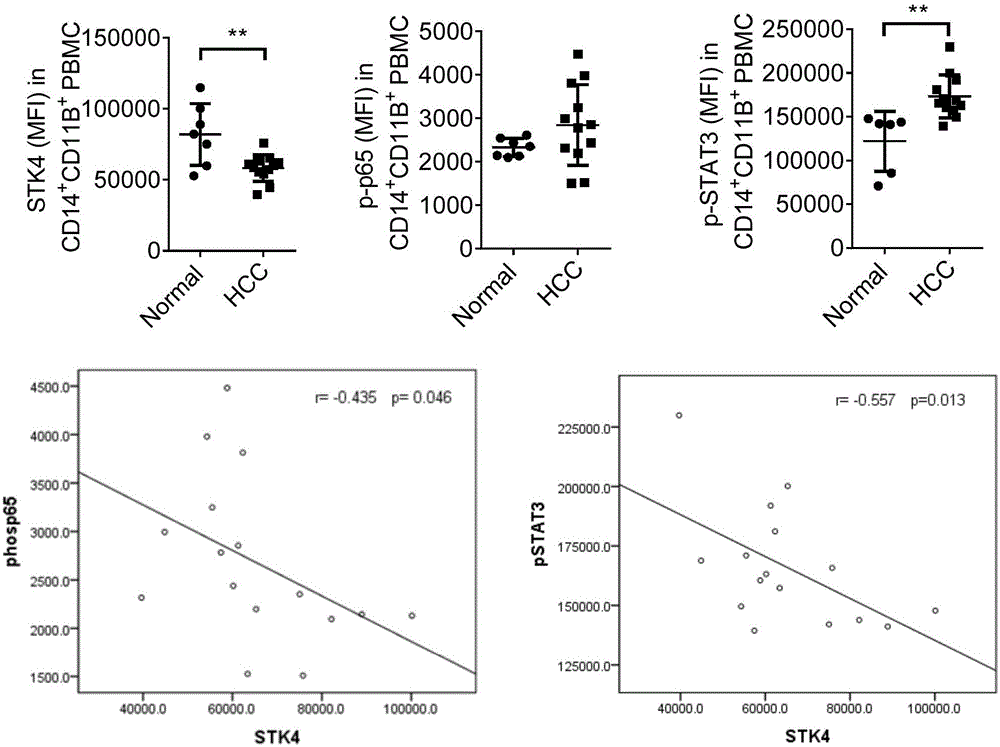
[0068] Example 3. Combined detection of STK4, p-p65 and p-STAT3 levels in peripheral blood mononuclear cells of patients with liver cancer, application in individualized treatment

[0069] Peripheral blood mononuclear cells were collected from 7 normal healthy people and 12 liver cancer patients. 3ml of blood collected from normal healthy people or liver cancer patients was collected by anticoagulant tube, separated by Ficoll, and the middle layer mononuclear cells were collected, and CD14 was detected by FACS flow cytometry + CD11b + Contents of STK4 and p-p65 and p-STAT3 in peripheral blood mononuclear cells.

[0070] The result is as image 3 , in patients with liver cancer CD14 + CD11b + In peripheral blood mononuclear cells, CD14 was detected by flow cytometry + CD11b + The expression level of STK4 in peripheral blood mononuclear cells is lower than that of normal people. On the contrary, both phos-p65 and phos-STAT3 were higher than normal.

[0071] Moreover, usi...


PUM


Abstract

The invention relates to a combined diagnostic marker for inflammation-associated HCC and an application thereof. Combined application of an STK4 protein in a peripheral blood monocyte or plasma, phosphorylated p65 (p-p65) and phosphorylated STAT3 (p-STAT3) in the peripheral blood monocyte and an IL-6 protein in the plasma as the marker for inflammation-associated HCC is provided for the first time, and a theoretical basis and a simpler detection method are provided for fast and early diagnosis and monitoring of HCC.

Description

technical field [0001] The present invention belongs to the field of disease diagnosis, and more specifically, the present invention relates to the early large-scale, specific screening of diagnostic markers of inflammation-related liver cancer and the application of individualized treatment plans for targets with abnormal expression of markers. Background technique [0002] At present, cancer is still one of the most harmful diseases to human health in the world, and it is also one of the most difficult diseases to overcome. Liver cancer is one of the most common malignant tumors in my country. Clinically diagnosed patients with liver cancer are generally in the middle and advanced stages, with severe liver function damage, and even after surgery and chemotherapy, the survival rate is very low. Therefore, early detection and early treatment are crucial. In recent years, with the rapid development of research in the field of life sciences, the detection of tumor markers in ...

Claims


Application Information

Patent Timeline
Patent Type & Authority Applications(China)
IPC IPC(8): G01N33/574G01N33/68
Inventor 王红艳李伟芸魏滨
Owner CENT FOR EXCELLENCE IN MOLECULAR CELL SCI CHINESE ACAD OF SCI
Why Eureka
Try Eureka