
Trachea cannula tooth cushion

A technique for endotracheal intubation and tooth pads, which is applied in the field of endotracheal intubation and tooth pads, can solve the problems of complicated fixation methods of tooth pads, suffocation, lung infection, and injury to the upper jaw, etc., so as to facilitate secretion processing and prevent hypostatic pneumonia. , easy to install and remove

Inactive Publication Date: 2017-06-20
THE FIRST AFFILIATED HOSPITAL OF SOOCHOW UNIV
View PDF8 Cites 14 Cited by
  • Summary
  • Abstract
  • Description
  • Claims
  • Application Information

AI Technical Summary

Problems solved by technology

[0003] Existing endotracheal intubation tooth pads have many defects. When a hard plastic tubular mouthpiece is used as a tooth pad, since the mouthpiece is made of hard plastic, it will cause damage to the patient's teeth, gums, oral mucosa, lips, etc. during use. Damage, comatose patients gnaw their teeth and cause tooth damage, and the tube that penetrates into the mouth can also damage the palate. For patients with missing teeth and exposed gums, the damage caused by using the existing tubular dental block is greater
The fixing method of the existing tooth pad is relatively complicated, and at the same time, the firmness is low, and it is easy to block the scale of the endotracheal intubation, which is not conducive to the observation of medical staff
Pasting and fixing with adhesive tape will have adverse effects on the skin of the patient's mouth and cheeks, and some patients will have allergic reactions
The existing fixation method of tracheal intubation is not conducive to the observation and nursing operation of the oral mucosa of the patient by the medical staff, and the repeated removal of the adhesive tape is inefficient and easily causes discomfort to the patient
Patients with endotracheal intubation have a lot of secretions such as sputum and saliva in the oral cavity, which are easy to breed bacteria. In severe cases, they will flow into the airway and cause suffocation and lung infection. The existing technology generally adopts the method of regular suction for treatment. The method cannot guarantee aseptic operation, which is easy to cause infection

Method used


Image

  • Trachea cannula tooth cushion
  • Trachea cannula tooth cushion

Examples


Embodiment Construction

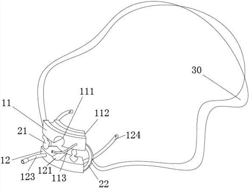
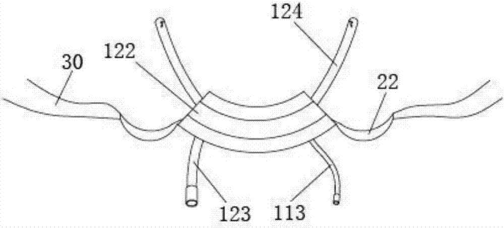
[0019] Such as figure 1 with figure 2 Shown is a schematic structural view of a tooth pad for endotracheal intubation of the present invention, which includes an upper tooth pad 11, a lower tooth pad 12, two air bags 21, two arcuate walls 22 and a fixed Belt 30, the upper dental pad 11 and the lower dental pad 12 are connected through the two arcuate walls 22 and the air bag 21, and the two arcuate walls 22 connect the upper dental pad 11 and the lower dental pad 12 , and the arcuate wall 22 connects the corresponding two ends of the upper dental pad 11 and the lower dental pad 12, and the two ends of the fixing belt 30 are respectively connected with the two arcuate walls 22.

[0020] Further, the upper dental pad 11 is arc-shaped, the upper dental pad 11 has an upper slot 111 and an upper dental groove 112, the upper slot 111 is located at the bottom of the upper dental pad 11, and the upper The clamping slot 111 is cylindrically recessed in the upper dental pad 11, and t...


PUM


Abstract

The invention discloses a trachea cannula tooth cushion. The trachea cannula tooth cushion comprises an upper tooth cushion body and a lower tooth cushion body. The upper and lower tooth cushion are connected through arch-shaped arms and air bags on the two sides, and a trachea cannula is placed between the upper and lower tooth cushions, penetrates through the upper and lower tooth cushions and enters the airway of a patient. According to the conditions of the patient, the oral cavity of the patient can be expanded by inflating the air bags, and operation is convenient. The tooth cushion is in a U shape, conforms to the oral cavity structure of the human body, and can easily protect teeth, gingivae and mouth mucosa of the patient. Sputum suction pipes are installed on the tooth cushion, secreta can be sucked out, and water or medicine can also be injected. The tooth cushion is fixed through a flat fixing band, fixing is firm, and the comfort degree of the patient is improved.

Description

technical field [0001] The invention relates to the field of clinical nursing medical equipment, and more precisely relates to a tooth pad for tracheal intubation. Background technique [0002] In the field of clinical nursing, especially in the rescue and treatment of critically ill patients, tracheal intubation technology is often required. Endotracheal intubation refers to the technique of inserting a special endotracheal tube into the trachea through the glottis. good condition. Most critically ill patients who use endotracheal intubation use ventilator-assisted ventilation, and the time of intubation needs to be determined according to the change of the condition, ranging from a few days to several months. to patient recovery and comfort. Some patients who need endotracheal intubation are in a coma state, and their trismus cannot cooperate with observation and treatment. Some patients may involuntarily move their lips, teeth and tongue due to a foreign body sensation...

Claims


Application Information

Patent Timeline
IPC IPC(8): A61M16/04
CPCA61M16/0493A61M16/0497A61M2210/0637
Inventor 袁德敬陆晓燕李俊杰
Owner THE FIRST AFFILIATED HOSPITAL OF SOOCHOW UNIV
Learn More