Retinal vessel segmentation method based on combination of deep learning and traditional method

A retinal blood vessel and deep learning technology, applied in the field of retinal blood vessel segmentation, can solve problems such as low calculation efficiency, classification failure, and large storage overhead, and achieve the goals of improving robustness and accuracy, wide applicability, and reducing feature redundancy Effect

Active Publication Date: 2017-07-04
BEIJING UNIV OF TECH
View PDF4 Cites 48 Cited by
  • Summary
  • Abstract
  • Description
  • Claims
  • Application Information

AI Technical Summary

Problems solved by technology

However, classifying each pixel rarely involves global information, so that the classification fails in the case of local lesions; secondly, each image has at least hundreds of thousands of pixels, and if it is judged one by one, the storage overhead will be large, and the calculation low efficiency

Method used


Image

  • Retinal vessel segmentation method based on combination of deep learning and traditional method
  • Retinal vessel segmentation method based on combination of deep learning and traditional method
  • Retinal vessel segmentation method based on combination of deep learning and traditional method

Examples


Embodiment Construction

[0040] Describe in detail below in conjunction with accompanying drawing:

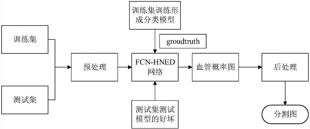
[0041] The technical block diagram of the present invention is as figure 1 shown. The specific implementation steps are as follows:

[0042] 1. Pretreatment

[0043] The same preprocessing is performed on each retinal fundus image whether it is a training set or a test set.

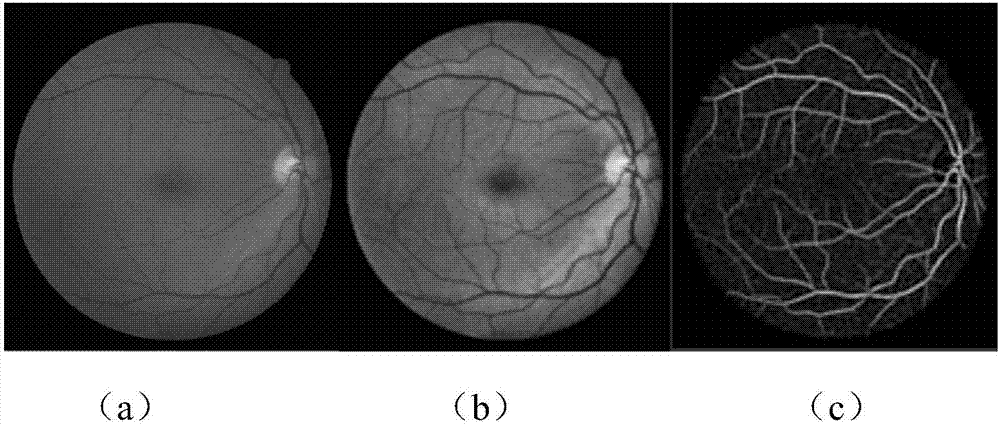
[0044] 1) Extract the green channel with relatively high contrast in the RGB three channels of the color retinal image. Secondly, due to problems such as shooting angles, the brightness of the collected retinal fundus images is often uneven, or the lesion area is difficult to distinguish from the background due to problems such as too bright or too dark and low contrast in the image. Therefore, we Perform normalization processing, and then perform CLAHE processing on the normalized retinal image to improve the quality of the retinal fundus image, balance the brightness of the fundus image, and make it more suitable for subsequ...


PUM


Abstract

The invention discloses a retinal vessel segmentation method based on combination of deep learning and a traditional method and relates to the fields of computer vision and mode recognition. According to the method, two grayscale images are both used as training samples of a network, corresponding data amplification, including elastic deformation, smooth filtering, etc., is done against the problem of less retinal image data, and wide applicability of the method is improved. According to the method, an FCN-HNED retinal vessel segmentation deep network is constructed, an autonomous learning process is realized to a great extent through the network, convolutional features of a whole image can be shared, feature redundancy can be reduced, the category of multiple pixels can be recovered from the abstract features, a CLAHE graph and a gauss matched filtering graph of the retinal vessel image are input into the network, an obtained vessel segmentation graph is subjected to weighted average, and therefore a better and more intact retinal vessel segmentation probability graph is obtained. Through the processing mode, the robustness and accuracy of vessel segmentation are improved to a great extent.

Description

technical field [0001] The invention relates to the fields of computer vision and pattern recognition, and is a retinal blood vessel segmentation method based on the combination of deep learning and traditional methods. Background technique [0002] Fundus imaging can be used to determine whether there is an abnormality through retinal imaging, and the observation of retinal blood vessels is very important. Diseases such as glaucoma, cataract, and diabetes can all cause lesions in the blood vessels of the retinal fundus. The number of patients with retinopathy is increasing year by year, and if it is not treated in time, it usually leads to great pain and even blindness in patients with long-term suffering from these diseases. However, at present, retinopathy is manually diagnosed by a specialist. The specialist first manually marks the blood vessels on the patient's fundus image, and then measures the required blood vessel diameter, bifurcation angle and other related para...

Claims


Application Information

Patent Timeline
Patent Type & Authority Applications(China)
IPC IPC(8): G06T7/00G06T7/12
CPCG06T7/0012G06T2207/20081G06T2207/20084G06T2207/30041
Inventor 蔡轶珩高旭蓉邱长炎崔益泽王雪艳孔欣然
Owner BEIJING UNIV OF TECH
Why Eureka
Try Eureka