Multiple-degree-of-freedom animal cone-beam CT imaging system

A CT imaging, degree-of-freedom technology, applied in medical science, clinical application of radiological diagnosis, control of radiological diagnostic equipment, etc., to achieve the effect of high application value and high flexibility

Pending Publication Date: 2017-09-01
ZHEJIANG UNIV
View PDF6 Cites 13 Cited by
  • Summary
  • Abstract
  • Description
  • Claims
  • Application Information

AI Technical Summary

Problems solved by technology

[0005] Aiming at the above-mentioned deficiencies in the prior art, the present invention provides a multi-degree-of-freedom animal cone-beam CT imaging system, which solves the drawbacks of traditional cone-beam CT imaging systems that can only translate and scan multiple times for larger imaging positions to cover the entire lesion , with the advantages of multiple scanning methods, wide scanning range and adjustable imaging parameters

Method used


Image

  • Multiple-degree-of-freedom animal cone-beam CT imaging system
  • Multiple-degree-of-freedom animal cone-beam CT imaging system
  • Multiple-degree-of-freedom animal cone-beam CT imaging system

Examples


Embodiment Construction

[0016] The present invention will be further described in detail below in conjunction with the accompanying drawings and specific embodiments.

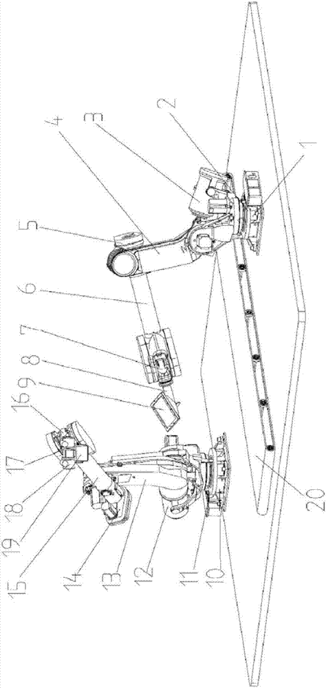
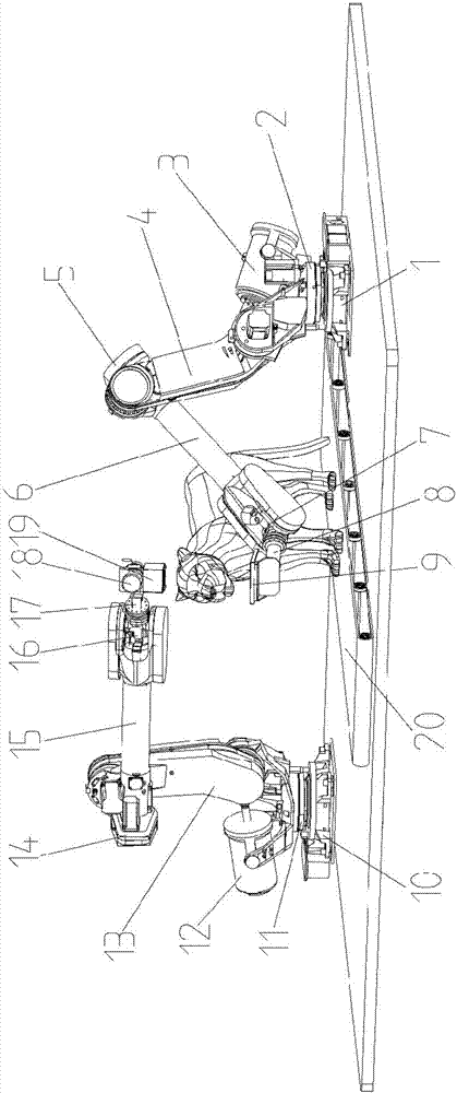
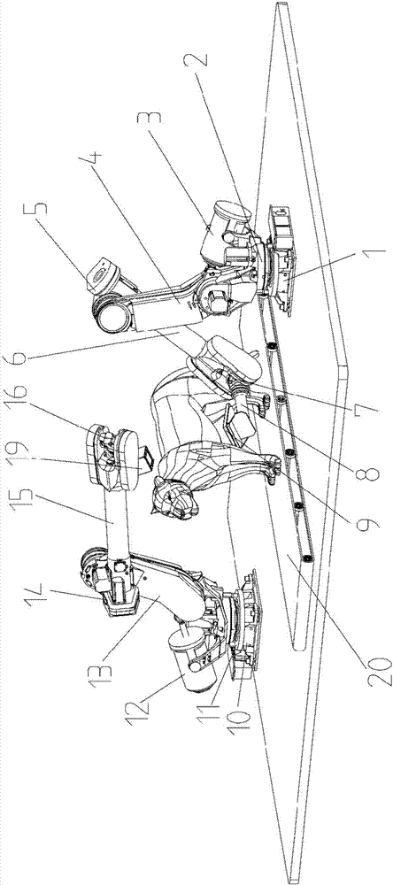
[0017] like Figure 1 ~ Figure 3 As shown, the present invention provides a multi-degree-of-freedom animal cone-beam CT imaging system, including a first six-axis robot arm with a detector 9 fixed at the end, a second six-axis robot arm with a tube 18 fixed at the end, and a synchronous conveyor belt 20 , wherein the first six-axis robot arm and the second six-axis robot arm are symmetrically installed on the left and right sides of the synchronous conveyor belt 20 .

[0018] The first six-axis mechanical arm includes: a first base 1, a first rotating platform 2, a first hydraulic cylinder 3, a first mechanical arm 4, a first rotating support 5, a second mechanical arm 6, and a first end motor 7. The detector bracket 8 and the detector 9, wherein: the first base 1 is fixed on the left side of the synchronous conveyor belt 20, the fir...


PUM


Abstract

The invention discloses a multiple-degree-of-freedom animal cone-beam CT imaging system. The system includes a first six-shaft mechanical arm, a second six-shaft mechanical arm and a synchronous conveyor belt, wherein the tail end of the first six-shaft mechanical arm is fixedly provided with a detector, and the tail end of the second six-shaft mechanical arm is fixedly provided with a ball pipe; the first six-shaft mechanical arm and the second six-shaft mechanical arm are symmetrically installed on the two sides of the synchronous conveyor belt, and the first six-shaft mechanical arm and the second six-shaft mechanical arm can achieve six-degree-of-freedom space movement. The system is high in flexibility, and for animals different in size and shape, a good scanning space and posture can be obtained through the position adjustment of the two six-shaft mechanical arms. The system has the non-coplanar scanning capability, and has a high application value in the field of high-end medical image guidance, such as precise intraoperative navigation. Since the position of the detector can be deviated, a large imaging region can be obtained in a half-fan-shaped mode when an imaging object is larger.

Description

technical field [0001] The invention belongs to the technical field of medical devices, in particular to a multi-degree-of-freedom animal cone-beam CT imaging system. Background technique [0002] Traditional cone-beam CT systems have limited scanning trajectories, and usually can only scan in a circular trajectory in a single scan, so the coverage of the longitudinal space is small. Therefore, in the clinical treatment of head and neck tumors, the "step-exposure" mode is often adopted, that is, after completing a circular trajectory scan, move to the next body position to continue scanning, and scan successively to cover the entire lesion. During this process, the radiation dose will increase significantly, and the subsequent image stitching process is also relatively cumbersome. [0003] In addition, due to the lack of non-coplanar scanning capability, traditional cone-beam CT imaging is difficult to image-guidance during stereotaxic radiosurgery, reducing the therapeutic...

Claims


Application Information

Patent Timeline
Patent Type & Authority Applications(China)
IPC IPC(8): A61B6/03A61B6/14
CPCA61B6/032A61B6/14A61B6/4085A61B6/4452A61B6/4458A61B6/508A61B6/54A61B6/587A61B2503/40
Inventor 牛田野杨春林刘桥王静吴式琇
Owner ZHEJIANG UNIV
Why Eureka
Try Eureka