Multi-modal image fusion surgical system and navigation method

An image fusion and surgical system technology, applied in the field of image processing systems, can solve problems such as radiation damage to doctors and patients, high price and maintenance costs of angiography machines, insufficient contrast to reflect the details of lesions, etc., to achieve easy upgrades and improve visualization experience , easy maintenance

Inactive Publication Date: 2017-10-10
卡本(深圳)医疗科技有限公司
View PDF5 Cites 2 Cited by
  • Summary
  • Abstract
  • Description
  • Claims
  • Application Information

AI Technical Summary

Problems solved by technology

The advantage of X-ray assisted therapy technology is that it is real-time, and the fluoroscopic image clearly reflects the anatomical characteristics of the human body; the disadvantage is that it has radiation damage to doctors and patients, and the contrast is not enough to reflect the details of the lesion, such as the scar of liver cancer. The price and maintenance cost of angiography machine are relatively high. high

Method used


Image

  • Multi-modal image fusion surgical system and navigation method

Examples


Embodiment Construction

[0028] The present invention will be further described below in conjunction with the accompanying drawings and embodiments.

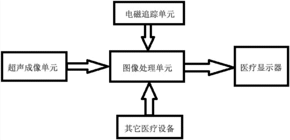
[0029] A multimodal image fusion surgery system, comprising: an ultrasound imaging unit, an image processing unit, an electromagnetic tracking unit, and a medical display; the output terminals of the ultrasound imaging unit and the electromagnetic tracking unit are connected to the input terminals of the image processing unit, and the The output end of the image processing unit is connected to a medical monitor.

[0030] Wherein, the ultrasonic imaging unit includes a medical ultrasonic probe, an ultrasonic imaging system and an ultrasonic data interface; the input end of the ultrasonic imaging system is connected to the output end of the medical ultrasonic imaging probe, and the output end of the ultrasonic imaging system is connected to the image processing unit through the ultrasonic data output interface. input.

[0031] The electromagnetic trackin...


PUM


Abstract

A multimodal image fusion surgery system and navigation method, the system includes: an ultrasound imaging unit, an image processing unit, an electromagnetic tracking unit and a medical display; the output ends of the ultrasound imaging unit and the electromagnetic tracking unit are connected to the image processing unit The input end, the output end of the image processing unit is connected to a medical display. After using the present invention, the fusion of different modal image data of the same lesion of the same patient can be completed, and a radiation-free, high-efficiency, multi-view navigation product can be provided for surgery.

Description

technical field [0001] The present invention relates to an image processing system, in particular to an operation system and a navigation method based on multimodal medical image fusion. Background technique [0002] In the era of minimally invasive surgery, X-ray surgical navigation equipment such as angiography machine and gastrointestinal machine have become the main surgical tools. Use the fluoroscopy function of X-ray equipment to navigate the probes, guide wires, catheters, etc. entering the human body, and assist doctors to complete the surgical treatment of the lesion. The advantage of X-ray assisted therapy technology is that it is real-time, and the fluoroscopic image clearly reflects the anatomical characteristics of the human body; the disadvantage is that it has radiation damage to doctors and patients, and the contrast is not enough to reflect the details of the lesion, such as the scar of liver cancer. The price and maintenance cost of angiography machine are ...

Claims


Application Information

Patent Timeline
Patent Type & Authority Applications(China)
IPC IPC(8): A61B34/20A61B34/10
CPCA61B34/20A61B34/10A61B2034/107A61B2034/2051A61B2034/2063A61B2034/2065
Inventor 王晓芳
Owner 卡本(深圳)医疗科技有限公司
Why Eureka
Try Eureka