SAR value determination method of magnetic resonance imaging and magnetic resonance imaging device

A magnetic resonance imaging and determination method technology, which is applied in magnetic resonance measurement, material analysis through resonance, magnetic property measurement, etc., can solve the problems of unfixed position and inability to obtain SAR values ​​accurately, so as to ensure safety and improve scanning speed and image quality, human exposure estimation model accurate effect

Active Publication Date: 2017-10-24
SHANGHAI UNITED IMAGING HEALTHCARE
View PDF7 Cites 6 Cited by
  • Summary
  • Abstract
  • Description
  • Claims
  • Application Information

AI Technical Summary

Problems solved by technology

However, in some special scans such as knee joint scans or infant scans, the position on the scanning table is not fixed, and the position of the human body in the scannin

Method used


Image

  • SAR value determination method of magnetic resonance imaging and magnetic resonance imaging device
  • SAR value determination method of magnetic resonance imaging and magnetic resonance imaging device
  • SAR value determination method of magnetic resonance imaging and magnetic resonance imaging device

Examples


Embodiment Construction

[0041] In order to make the above objects, features and advantages of the present invention more obvious and comprehensible, specific implementations of the present invention will be described in detail below in conjunction with the accompanying drawings and embodiments.

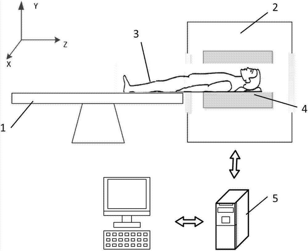
[0042] A magnetic resonance imaging system usually includes a magnet with a certain aperture, a transmitting coil for transmitting radio frequency signals and a receiving coil for receiving magnetic resonance signals, a gradient coil for spatially positioning the magnetic resonance signals, and a sensor for generating scan sequences. Pulse generator and control system. The MRI system works through an operator (clinician) controlling a console connected to the control system, which may include a keyboard or other input device, a control panel, and a display to enter commands and display generated images.

[0043] figure 1 is a schematic diagram of the structure of the magnetic resonance imaging device. Durin...


PUM


Abstract

The invention discloses an SAR value determination method of magnetic resonance imaging. The method comprises the steps that patients are placed in a scanning table of a magnetic resonance imaging system, the scanning table is moved, so that the patients are moved toward a scanning area, the quality of body parts, located on the scanning area, of the patients, is obtained, and an SAR value of parts of bodies is determined according to the quality of the body parts which are located on the scanning area; the process of obtaining the quality of the body parts which are located on the scanning area comprises the steps that a frequency domain characteristic curve of a body part outline on the selected level position is obtained, the parts to be imaged which is located on the scanning area are identified according to the distribution of a signal area of the characteristic curve, and the position of the parts to be imaged is determined according to the position of the scanning table; according to the weight and the height of the patients, and the position of the parts to be imaged, the quality of body parts which enter the scanning area is obtained. According to the SAR value determination method, a precise human body exposure estimation model can be built, and the scanning speed and the imaging quality are improved. Furthermore, the invention further provides a magnetic resonance imaging device.

Description

【Technical field】 [0001] The invention relates to the technical field of magnetic resonance imaging, in particular to a related technology capable of accurately monitoring the local specific absorption rate in the scanning area of ​​the magnetic resonance imaging. 【Background technique】 [0002] Magnetic resonance imaging (MRI, magnetic resonance imaging) is a high-tech imaging based on the performance characteristics of biological hydrogen nuclei in a magnetic field. The physical basis of magnetic resonance imaging is the resonance phenomenon related to material magnetism and magnetic field, that is, radio frequency waves The angular momentum has the magnetic moment and the resonant properties exhibited by the system interacting in an external magnetic field. The clinical application in the central nervous system has become the gold standard for disease diagnosis, and it is also unique in the diagnosis of bone, joint and soft tissue lesions. In recent years, ultra-high-fie...

Claims


Application Information

Patent Timeline
IPC IPC(8): A61B5/055G01R33/20
CPCA61B5/055G01R33/20
Inventor 邢晓聪
Owner SHANGHAI UNITED IMAGING HEALTHCARE
Why Eureka
Try Eureka