Eureka AIR delivers breakthrough ideas for toughest innovation challenges, trusted by R&D personnel around the world.

Method for constructing gastrointestinal stromal tumor malignant potential classifying model based on radiomics

A technology for gastrointestinal stromal tumor and classification model, which is applied in the fields of imaging, computer-aided medicine, and oncology to improve the correlation, reduce the error rate, and avoid damage.

Inactive Publication Date: 2017-11-21
NANFANG HOSPITAL OF SOUTHERN MEDICAL UNIV
View PDF0 Cites 6 Cited by
  • Summary
  • Abstract
  • Description
  • Claims
  • Application Information

AI Technical Summary

Problems solved by technology

In summary, although the existing GIST classification methods can provide some clues and directions for clinicians to diagnose and treat, there is still a lot of room for improvement and improvement.

Method used


Image

  • Method for constructing gastrointestinal stromal tumor malignant potential classifying model based on radiomics
  • Method for constructing gastrointestinal stromal tumor malignant potential classifying model based on radiomics
  • Method for constructing gastrointestinal stromal tumor malignant potential classifying model based on radiomics

Examples


Embodiment Construction

[0027] 1. Data collection:

[0028] (1) Develop criteria such as (a) GIST patients who have not received adjuvant imatinib therapy (b) GIST after complete resection (c) less than 15 days of abdominal enhanced CT before surgery. Exclude the influence of other factors on this experiment.

[0029] (2) Obtain abdominal CT images of GIST patients who have not undergone adjuvant imatinib treatment, and divide them into training group and prediction group.

[0030] (3) To test whether the differences between the cases regarding age, gender, tumor primary site, histological grade and other potential influencing factors are statistically significant.

[0031] 2. Extract radiomic features:

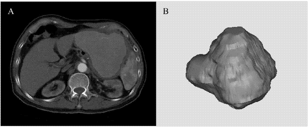
[0032] Use ITK-SNAP software to outline the ROI, outline the tumor contour layer by layer, and then perform 3D volume reconstruction of the 2D ROI to generate VOI (such as figure 1 Shown), using Matlab 2014b software to extract feature data, including texture features and non-texture features.

[0033] Note: ...


PUM


Abstract

The invention is a method of tumorology, imaging, and computer -assisted medicine, involving a method of building a model of malignant potential classification models based on radiological gastric and intestinal tumor.The construction method described in the present invention includes: data collection: abdominal enhancement phase of thin -layer CT image collection; extracting radiation group characteristics; statistical analysis; feature selection and radiation group characteristics model construction; radiology feature model verification and calibration.The method built by the method described by the present invention can classify the vicious potential classification model of gastrointestinal gastrointestinal tumor tumor tumor, which can accurately classify the GIST with different malignant potential, which is a non -invasive technology without increasing the additional cost of patients. Operations operation. Operations.Simple, convenient for clinical promotion.

Description

Technical field [0001] The invention belongs to the fields of oncology, imaging and computer-aided medicine, and relates to a method for constructing a gastrointestinal stromal tumor (Gastrointestinal Stromal Tumors, GIST) classification model based on radiomics. Background technique [0002] Gastrointestinal Stromal Tumors (GIST) are a type of tumors that originate from the gastrointestinal stromal tissues and account for most of the gastrointestinal mesenchymal tumors. Immunohistochemical detection usually expresses CD117, showing that Cajal cells differentiate, and most cases have c-kit or PDGFRA activating mutations. In recent years, the incidence of GIST in China has been rising rapidly. The only way to cure GIST is surgical resection, but the recurrence and metastasis rate of high-risk GIST patients can be as high as 55% to 90%. The adjuvant therapy of small-molecule targeted drugs-imatinib for tumors with high recurrence potential can significantly reduce the probability...

Claims


Application Information

Patent Timeline
IPC IPC(8): G06F19/00G06T7/00G06T7/40
CPCG06T7/40G06T7/0012G06T2207/30092
Inventor 陈韬李国新
Owner NANFANG HOSPITAL OF SOUTHERN MEDICAL UNIV
Why Eureka
Eureka Blog
Learn More