
PET image reconstruction method and PET imaging device

An image reconstruction and image technology, which is applied in the field of medical imaging, can solve problems such as blurred images, no obvious improvement in the definition of organ boundaries, and reduced accuracy of lesion areas, so as to achieve the effect of improving quality

Active Publication Date: 2017-12-29
SHENYANG INTELLIGENT NEUCLEAR MEDICAL TECH CO LTD
View PDF5 Cites 10 Cited by
  • Summary
  • Abstract
  • Description
  • Claims
  • Application Information

AI Technical Summary

Problems solved by technology

[0003] However, the joint potential function adopted by the joint prior model based on anatomical function only introduces a simple quadratic penalty term, which will cause the final PET reconstruction image to become blurred while improving the uniformity of the image, and the resulting The above joint potential function only calculates the quadratic potential function between MRI image pixels and adjacent pixels, so that the clarity of organ boundaries in PET reconstruction images is not significantly improved compared with traditional reconstruction results
In addition, since MRI images reflect anatomical information and have high image resolution and spatial texture information, the metabolic regions of organs (that is, the regions within organ boundaries) may not be evenly distributed in MRI images. In this case When the spatial texture information in the MRI image is introduced into the PET reconstruction image, the uniformity of the organ metabolic area in the PET reconstruction image will become worse, that is, the introduced MRI texture information will reduce the determination of the lesion area in the PET reconstruction image. correctness

Method used


Image

  • PET image reconstruction method and PET imaging device
  • PET image reconstruction method and PET imaging device
  • PET image reconstruction method and PET imaging device

Examples


Embodiment Construction

[0052] In order to make the purposes, technical solutions and advantages of the embodiments of the present application clearer, the technical solutions in the embodiments of the present application will be clearly and completely described below in conjunction with the drawings in the embodiments of the present application. Obviously, the described embodiments It is a part of the embodiments of this application, not all of them. Based on the embodiments in this application, all other embodiments obtained by persons of ordinary skill in the art without making creative efforts belong to the scope of protection of this application.

[0053] When the traditional iterative algorithm is simply used for PET image reconstruction, due to the insufficient count rate of the measured projection data and errors such as system noise, the reconstructed image contains more noise and the image uniformity is not ideal. In order to overcome this defect, the prior art provides a method for reconst...


PUM


Abstract

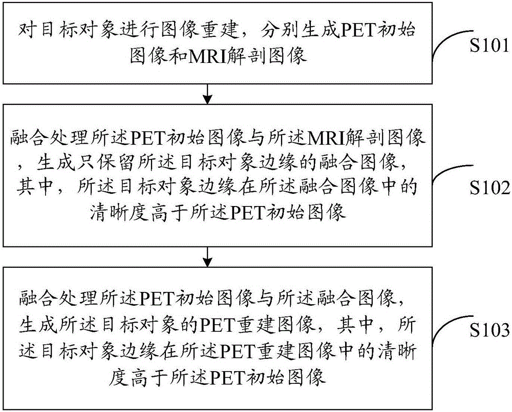
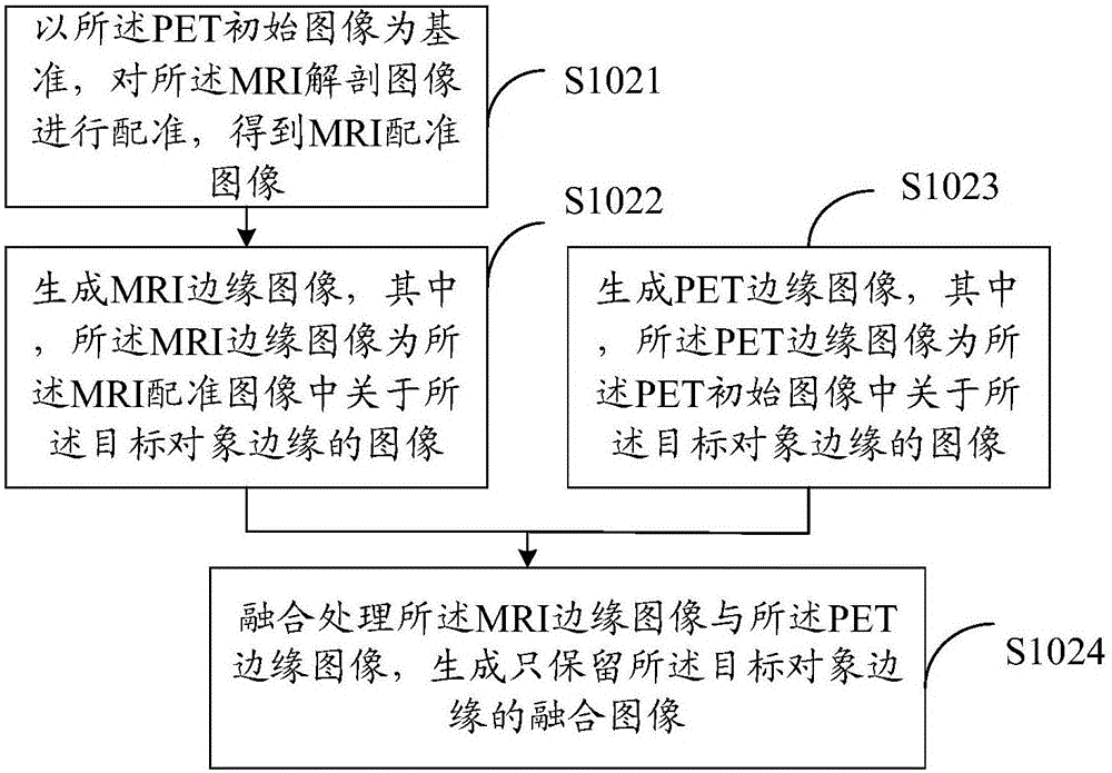
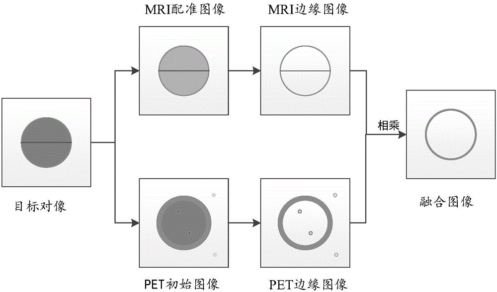
The application discloses a PET image reconstruction method and a PET imaging device. The method comprises steps of: performing image reconstruction on a target object to generate a PET initial image and an MRI anatomical image; fusing the PET initial image with the MRI anatomical image to generate a fusion image that retains only a target object edge, wherein the sharpness of the target object edge in the fusion image is higher than that of the PET initial image; fusing the PET initial image and the fusion image to generate a PET reconstruction image of the target object, wherein the sharpness of the target object edge in the PET reconstruction image is higher than that of the PET initial image. The PET image reconstruction method can improve the sharpness of the edge of the target object in the PET reconstruction image and ensure the uniformity of the metabolic region of the target object in the PET reconstruction image.

Description

technical field [0001] The present application relates to the technical field of medical imaging, in particular to a PET image reconstruction method and PET imaging equipment. Background technique [0002] Positron Emission Computed Tomography (PET) is a relatively advanced clinical examination imaging technology in the field of nuclear medicine. Multimodal medical images have been widely used in clinical diagnosis and treatment because they can provide more detailed image information, such as Magnetic Resonance Imaging (MRI) technology, which obtains anatomical images, and image characteristics High spatial texture information. According to research, there is a great correlation between MRI images and PET images, so that high-resolution MRI images can provide a large amount of prior information for PET image reconstruction. The experimental model was used for PET image reconstruction. [0003] However, the joint potential function adopted by the joint prior model based o...

Claims


Application Information

Patent Timeline
Patent Type & Authority Applications(China)
IPC IPC(8): G06T7/32G06T7/181
CPCG06T7/181G06T7/32G06T2207/10088G06T2207/20221G06T5/50G06T2207/20016G06T2207/20064G06T2207/20192G06T2207/30004G06T2207/10104G06T11/008G06T2211/464G06T5/70G06T7/337G06T7/13G06T2207/30096
Inventor 李运达孙智鹏刘勺连
Owner SHENYANG INTELLIGENT NEUCLEAR MEDICAL TECH CO LTD
Learn More