
Flap perforating branch artery locating method based on 3D printing reconstruction

A technology of 3D printing and positioning method, applied in the field of medical devices, can solve problems such as failure to find 3D printing, inability to accurately select acupoints, and troubles

Inactive Publication Date: 2018-01-23
岳春华
View PDF23 Cites 10 Cited by
  • Summary
  • Abstract
  • Description
  • Claims
  • Application Information

AI Technical Summary

Problems solved by technology

[0005] At present, the arteriovenous system inside the human body can only be displayed through radiology computer scans and angiography. Large blood vessels can also be reconstructed in 3D and printed out as a physical model, but the flap-perforating arteries belong to the microcirculatory system, with a diameter of only 0.5mm- 1mm, in the past, it was determined by autopsy. At present, no 3D printing method has been found, especially where the corresponding skin surface is. The surgical scope is simulated and selected by the doctor's experience, and there is no precise marking before the operation. Non-invasive means and tools for positioning, there are large errors and risks
[0006] More importantly, traditional acupuncture points also vary from person to person. Acupuncture requires precise acupoint selection and positioning before acupuncture can be performed. However, everyone has different acupoint locations. The acupoints of different people are only roughly the same, not They are completely consistent, so there will be a lot of trouble when positioning
At present, there are many methods on the market through infrared positioning or proportional measurement. Because they are all in vitro measurement methods and do not involve the essence of human organs, the results of the detection are the same as the selection of acupoints by the human eye, and it is impossible to accurately select acupoints.

Method used


Image

  • Flap perforating branch artery locating method based on 3D printing reconstruction
  • Flap perforating branch artery locating method based on 3D printing reconstruction
  • Flap perforating branch artery locating method based on 3D printing reconstruction

Examples


Embodiment Construction

[0040] The present invention will be further described below in conjunction with the accompanying drawings.

[0041] A flap perforating artery positioning method based on 3D printing and reconstruction, characterized in that the view and data are transmitted to the processing device through the DICOM images of human CT and MR, and then the processing device performs the following steps on the view and data:

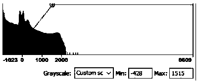
[0042] a. Adjust the view contrast: highlight the preprocessing part in the window, and modify the initial gray value of the overall image group. Due to the large individual differences in the image due to the use of machine models and other reasons, fine-tuning is required, such as figure 1 for the best contrast. (The CT value is represented by the scale HU) MIMICS only changes the contrast setting when adjusting the image, and will not affect the subsequent threshold segmentation.

[0043] b. Initial definition point selection and location: Sanyinjiao is where the Spl...


PUM


Abstract

The invention discloses a flap perforating branch artery locating method based on 3D printing reconstruction. The flap perforating branch artery locating method is characterized in that views and dataare transmitted to a processing device through DICOM images of human CT and MR, then aiming at the view and data, the processing device conducts the following processing steps that a, the views are compared and adjusted; b, the positions of selective points and determination points are firstly defined; c, a range domain is adjusted to determine the point-getting trend; d, point drawing tracing isconducted; e, a digitized 3D splitting model is manufactured; f, according to the model in the step e, 3D printing is conducted to obtain a physical model. The flap perforating branch artery locatingmethod has the advantages that the position of the flap perforating branch arteries can be accurately marked, at the same time, corresponding human organs are located and printed according to different parts, so that the surface of the 3D model has mark positions, corresponding to the skin, of the flap perforating branch arteries, the mark positions correspond to the human organs one to one, through model comparison, the positions, corresponding to the skin, of the human flap perforating branch arteries can be accurately judged, and the positions and shapes of hundreds of perforating branch arteries of all parts of the whole body can be accurately subjected to digitized virtual reconstruction and 3D physical printing.

Description

technical field [0001] The invention relates to the field of medical devices, in particular to a method for locating flap perforating arteries based on 3D printing reconstruction. Background technique [0002] On the anatomical map, there is a constant perforating branch of the posterior tibial artery about 5cm above the medial malleolus. The epithelial branch of the medial malleolus is the perforating branch of the intermuscular septum from the posterior tibial muscle group and the triceps muscle group of the calf. According to microsurgical anatomy, the three routes of anastomosis of the epithelial branch of the medial malleolus with the adjacent arterioles are the same as the three genital meridians. One is the anastomosis of the epithelial branch of the medial malleolus and the perforator of the posterior tibial artery (Jiaoxin, Taixi) under the deep fascial epithelium. The shape of the kidney meridian is the same. One is the anastomosis of the epithelial branch of the...

Claims


Application Information

Patent Timeline
Patent Type & Authority Applications(China)
IPC IPC(8): A61B34/10B33Y10/00B33Y50/00B33Y80/00
Inventor 岳春华李鹤虹
Owner 岳春华
Learn More