GBM multimodal magnetic resonance image segmentation method based on deep neural network

A technology of deep neural network and magnetic resonance image, which is applied in biological neural network model, neural learning method, image analysis, etc., can solve the problems of inability to speed up the segmentation speed and the explosion of network weights, and avoid the explosion of network weights , Speed ​​up the segmentation speed, and strengthen the effect of independent learning ability

Active Publication Date: 2018-03-06
ZHEJIANG CHINESE MEDICAL UNIVERSITY
View PDF6 Cites 30 Cited by
  • Summary
  • Abstract
  • Description
  • Claims
  • Application Information

AI Technical Summary

Problems solved by technology

[0010] The above invention still cannot speed up the segmentation sp

Method used


Image

  • GBM multimodal magnetic resonance image segmentation method based on deep neural network
  • GBM multimodal magnetic resonance image segmentation method based on deep neural network
  • GBM multimodal magnetic resonance image segmentation method based on deep neural network

Examples


Embodiment 1

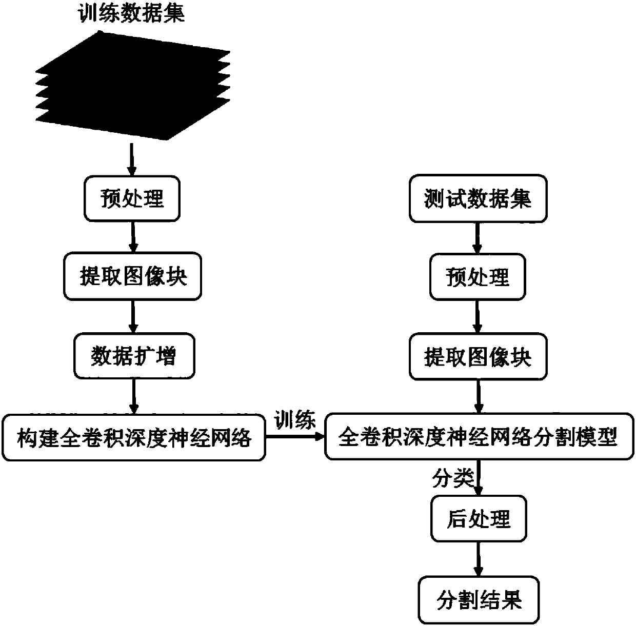
[0072] Embodiment 1, GBM multimodal magnetic resonance image segmentation method based on deep neural network, such as Figure 1-Figure 5 shown, including the following:

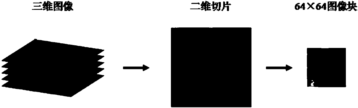
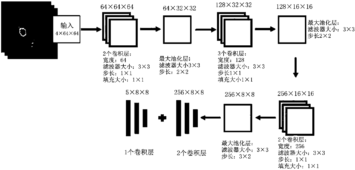
[0073] The GBM multimodal magnetic resonance image is segmented to obtain slice images, and each slice image is preprocessed to extract the image block, and then the trained deep neural network segmentation model is used to divide the 8× centered image block center voxel. All the voxels in the 8 area are classified, and the category of the voxel point is determined to be the normal tissue area of ​​the brain (hereinafter referred to as C 0 ), necrotic area (hereinafter referred to as C 1 ), edema area (hereinafter referred to as C 2 ), non-enhancing tumor area (hereinafter referred to as C 3 ) or enhanced tumor area (hereinafter referred to as C 4 ), so as to complete the segmentation of the four modality volume images of GBM, and the result of the post-processing is the final segmentation result of the ...


PUM


Abstract

The invention provides a GBM multimodal magnetic resonance image segmentation method based on a deep neural network. The method comprises the following steps that firstly each slice image of the collected GBM multimodal magnetic resonance image is preprocessed and then all the slice images are divided into training samples and test samples, and the slice images of the training samples are marked;then the training sample image blocks are extracted and the mean and the variance are standardized, and a training data asset is formed after data amplification; then one deep neural network is constructed and the deep neural network is trained by using the training data set so as to obtain a deep neural network segmentation model; and finally the slice image to be segmented is preprocessed and the image blocks are extracted, and the voxels are classified and post-processed by using the deep neural network segmentation model so that GBM multimodal magnetic resonance image segmentation can be realized. The high requirements of automatic diagnosis, surgical planning and prognosis for the detection and locating accuracy of the abnormal brain tissues and the surrounding normal structures can be met.

Description

technical field [0001] The invention relates to the fields of digital medical image processing and analysis and computer-aided diagnosis, in particular to a GBM multimodal magnetic resonance image segmentation method based on a deep neural network. Background technique [0002] Glioma is the most common primary brain tumor, which mainly occurs in adults, especially the elderly, and has three characteristics: high recurrence rate, high mortality rate and low cure rate. Relevant statistics show that more than half of the glioma patients are the most malignant GBM (Glioblastoma Multiforme, glioblastoma multiforme). Even with the most aggressive treatment, the median survival time of GBM patients is still less than 15 months, and the survival rate over 5 years is less than 5%. GBM presents a patch of heterogeneous tumor areas on multimodal magnetic resonance images. This area usually includes 4 parts: necrotic area, edema area, non-enhancing tumor area and enhancing tumor area...

Claims


Application Information

Patent Timeline
IPC IPC(8): G06T7/00G06T7/12G06T7/136G06N3/04G06N3/08
CPCG06T7/0012G06T7/12G06T7/136G06N3/08G06T2207/30016G06T2207/30096G06T2207/20081G06T2207/20084G06T2207/10088G06N3/045
Inventor 赖小波许茂盛徐小媚吕莉莉高卫红石磊
Owner ZHEJIANG CHINESE MEDICAL UNIVERSITY
Why Eureka
Try Eureka