Liver cancer image automatic segmentation method based on random forest and fuzzy clustering

A random forest and fuzzy clustering technology, applied in the field of medical image processing, can solve the problems of limited clinical application range, affecting the recognition effect, time-consuming and labor-consuming, etc., achieving fast calculation speed, good denoising effect, and wide application range Effect

Inactive Publication Date: 2018-03-27
NANJING UNIV OF SCI & TECH
View PDF9 Cites 24 Cited by
  • Summary
  • Abstract
  • Description
  • Claims
  • Application Information

AI Technical Summary

Problems solved by technology

Clinicians need to determine the location, size, and quantity of tumors when making surgical plans based on patients' CT images. If doctors manually segment lesions, it is very time-consuming and labor-intensive. Therefore, it is necessary to develop computer-aided automatic liver cancer segmentation algorithms.
The existing state-of-the-art technology (CN 105931224 A Method for Lesion Identification of Liver Plain Scan CT Image Based on Random Forest Algorithm) only has the recognition function and is only applicable to liver CT images in the plain scan period. The scope of clinical application is limited, and the lesion process can be identified from the image In the image preprocessing, the inherent noise in the image is not removed, which may affect the subsequent recognition effect

Method used


Image

  • Liver cancer image automatic segmentation method based on random forest and fuzzy clustering
  • Liver cancer image automatic segmentation method based on random forest and fuzzy clustering
  • Liver cancer image automatic segmentation method based on random forest and fuzzy clustering

Examples


Embodiment Construction

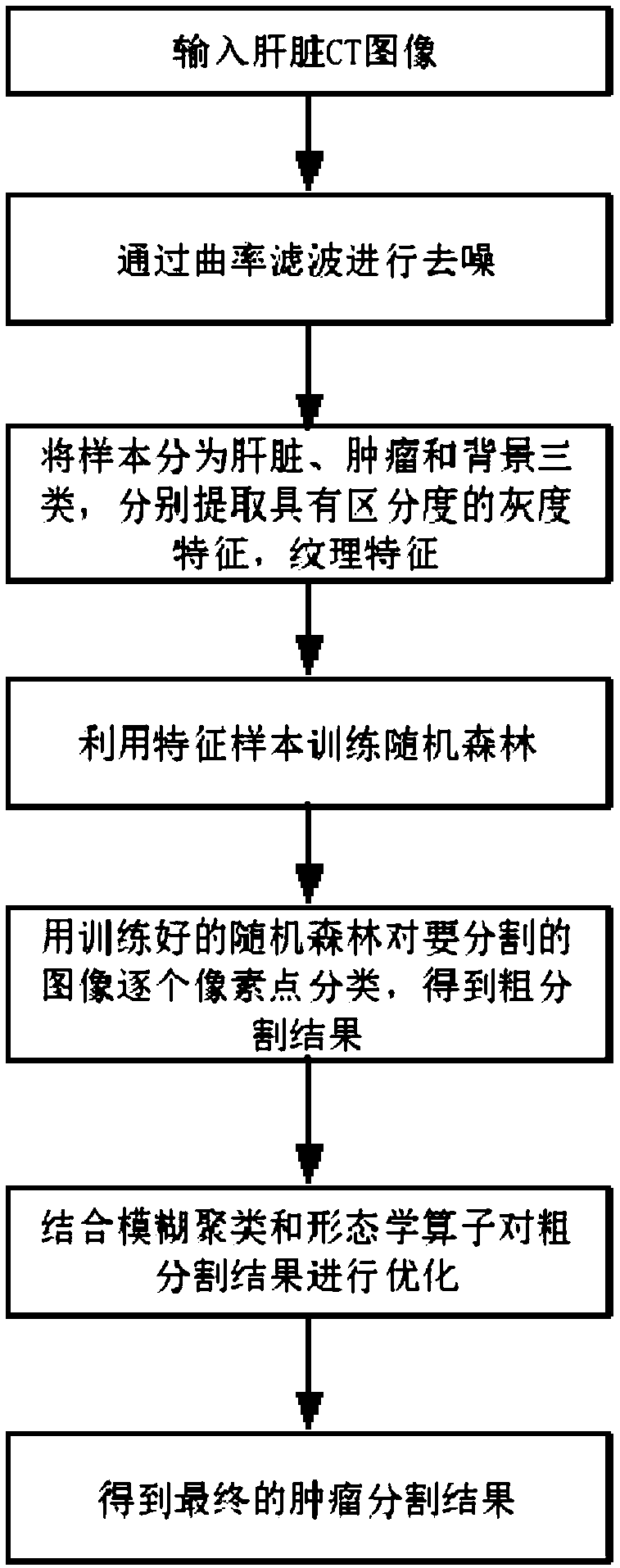
[0016] The present invention will be further described below in conjunction with accompanying drawing.

[0017] combine figure 1 , input a liver CT image with a size of 512×512×120, denoising by the following variational model

[0018]

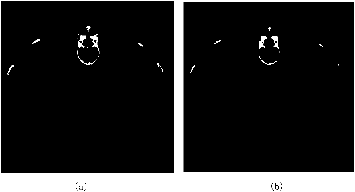
[0019] in represents a 2D image area, Represents the current denoised image, and treats the image as a three-dimensional surface, and represent the two principal curvatures of the surface, respectively, represents the Gaussian curvature. The model can be solved by the region decomposition method, and the denoising of the image can be realized after 15 iterations. The original image and the image after denoising are as follows: figure 2 .

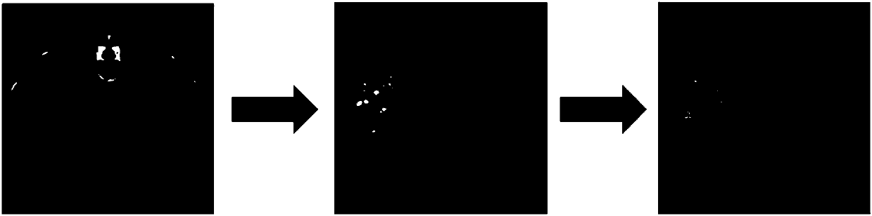
[0020] According to the gold standard of images in the training set (segmentation results of liver and tumor), the original images can be divided into three categories: liver, tumor and background. The grayscale features and texture features of the three types of images are extracted respectiv...


PUM


Abstract

The invention discloses a liver cancer CT image automatic segmentation method based on random forest and fuzzy clustering. The liver cancer CT image automatic segmentation method based on random forest and fuzzy clustering mainly has two processes: an image preprocessing process and a focus region segmentation process, and includes the following steps: in the preprocessing stage, removing the noise of a CT image through curvature filtering and at the same time enabling the edge of the image to be maintained unfuzzy; in the tumour image segmentation stage, training the random forest by extracting the character sample, classifying the pixel points one by one of the image to be segmented to obtain a coarse segmentation result, and then obtaining the final tumour segmentation result through fuzzy clustering and a morphological operator. The liver cancer CT image automatic segmentation method based on random forest and fuzzy clustering does not need adjusting parameters, can segment the liver cancer CT images in different period, has the advantages of being automatic, being high in accuracy and being wide in application, and has a wide application prospect in pre-operation programming and accurate treatment of liver cancer.

Description

technical field [0001] The invention belongs to the field of medical image processing, and in particular relates to the segmentation of liver tumors in CT (computed tomography) images. Background technique [0002] The liver is an organ in the human body that mainly has metabolic functions. It plays important roles in metabolism, storage of glycogen, and biotransformation in the body. However, the liver is also a site with a high incidence of lesions. Liver cancer can be divided into two categories: primary liver cancer and metastatic liver cancer, which are extremely harmful to the body and have various lesions. CT (computed tomography) technology is currently one of the most commonly used clinical imaging diagnostic techniques for liver cancer. Through CT scanning, doctors can obtain a series of secondary images of the liver in different periods (contrast period, venous period, arterial period and delayed period). Dimensional CT slices. Clinicians need to determine the l...

Claims


Application Information

Patent Timeline
Patent Type & Authority Applications(China)
IPC IPC(8): G06T7/10
CPCG06T2207/10081G06T2207/20081G06T2207/30096G06T7/10
Inventor 杨孝平刘芳马骏李渊强吴宇雳张梦璐
Owner NANJING UNIV OF SCI & TECH
Why Eureka
Try Eureka