Eureka AIR delivers breakthrough ideas for toughest innovation challenges, trusted by R&D personnel around the world.

A method and system for enhancing contrast-enhanced ultrasound images, and contrast-enhanced ultrasound imaging equipment

A contrast-enhanced ultrasound and contrast-enhancing technology, applied in ultrasonic/sonic/infrasound diagnosis, image enhancement, image data processing, etc., can solve the problems of submersion, the retention amplitude is quite or even smaller, and cannot be observed, so as to achieve the effect of contrast image enhancement.

Active Publication Date: 2020-11-24
BEIJING SHEN MINDRAY MEDICAL ELECTRONICS TECH RES INST +1
View PDF5 Cites 0 Cited by
  • Summary
  • Abstract
  • Description
  • Claims
  • Application Information

AI Technical Summary

Problems solved by technology

SNR is determined by the sensitivity of the probe and the system platform. A high SNR means that smaller microbubbles can be detected, and the smaller the microbubbles, the smaller the signal amplitude. The signal amplitude of some small microbubbles is equivalent to or even smaller than that of tissue residues. Therefore, even if the SNR is high, if the CTR is not high enough, the signal reflected by these small microbubbles will be submerged in the residual information of the tissue and cannot be observed in the contrast image

Method used


Image

  • A method and system for enhancing contrast-enhanced ultrasound images, and contrast-enhanced ultrasound imaging equipment
  • A method and system for enhancing contrast-enhanced ultrasound images, and contrast-enhanced ultrasound imaging equipment
  • A method and system for enhancing contrast-enhanced ultrasound images, and contrast-enhanced ultrasound imaging equipment

Examples


Embodiment Construction

[0041] The present invention will be further described in detail with reference to the drawings in the following specific embodiments.

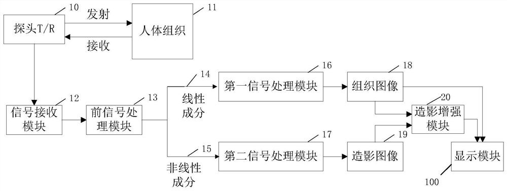
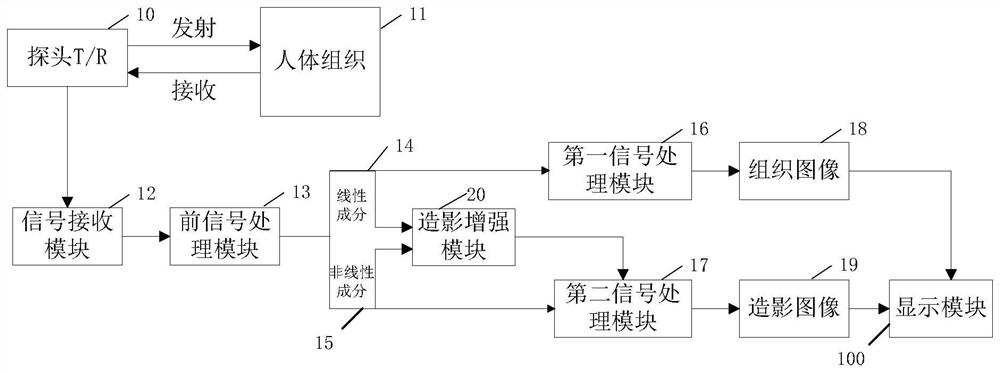
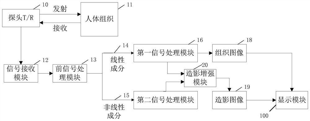
[0042] such as Figure 1 As shown, the contrast-enhanced ultrasound imaging device includes a probe 10, a front signal processing module 13, a first signal processing module 16, a second signal processing module 17, a tissue image generation module 18, a contrast-enhanced image generation module 19, a contrast-enhanced ultrasound image enhancement system 20 and a display module 100.

[0043] The probe 10 is used to transmit ultrasonic waves to the human tissue 11 and receive ultrasonic echoes reflected by the tissue 11, and the ultrasonic echoes can be stored in the signal receiving module 12.

[0044] The pre-signal processing module 13 is used to process the ultrasonic echo signal and extract useful information. The pre-signal processing module 13 mainly includes amplifiers, beam combiners and filters to extract more useful signals with differen...


PUM


Abstract

A method, system and ultrasound contrast imaging equipment for enhancing ultrasound contrast images. Based on nonlinear contrast information and linear tissue information obtained from ultrasound echo signals, nonlinear parameters are calculated to obtain nonlinear parameter distribution diagrams, respectively. The bipolar threshold divides the nonlinear parameter distribution map. Each division separates the contrast agent area and the tissue residual area. The contrast agent area is enhanced and the tissue residual area is suppressed, so that the area that absolutely belongs to the contrast agent can be processed. Enhance to a large extent, suppress areas that are definitely tissue residues to a large extent, enhance and suppress areas between contrast agents and tissue residues to a slightly weaker degree, and finally combine the results after two classifications and weight adjustments. Fusion processing enables the enhancement of contrast agents, suppression of contrast agents, and conservative treatment of residues between contrast agents and tissues to achieve ideal contrast image enhancement effects.

Description

Technical field [0001] The invention relates to contrast-enhanced ultrasound imaging equipment, in particular to a method and a system for enhancing contrast-enhanced ultrasound images. technical background [0002] Compared with conventional ultrasound imaging, contrast-enhanced ultrasound imaging can enhance the intensity of ultrasonic echo signal and show the tiny blood vessels that cannot be seen by conventional ultrasound. With the increasing requirements of clinical ultrasound examination, contrast-enhanced ultrasound imaging is more and more used in clinic, and it is more and more widely used to examine organs, and its position in clinical diagnosis is getting higher and higher. [0003] Contrast-enhanced ultrasound imaging is a kind of enhanced ultrasound imaging after injection of contrast agent, and contrast agent is an enhancer that can enhance the intensity of ultrasonic echo signal. There are many kinds of ultrasound contrast agents, and the most commonly used one is...

Claims


Application Information

Patent Timeline
IPC IPC(8): A61B8/00G06T5/00
CPCA61B8/00G06T5/90G06T2207/10132
Inventor 储霞唐杰桑茂栋朱磊张明博
Owner BEIJING SHEN MINDRAY MEDICAL ELECTRONICS TECH RES INST
Why Eureka
Eureka Blog
Learn More