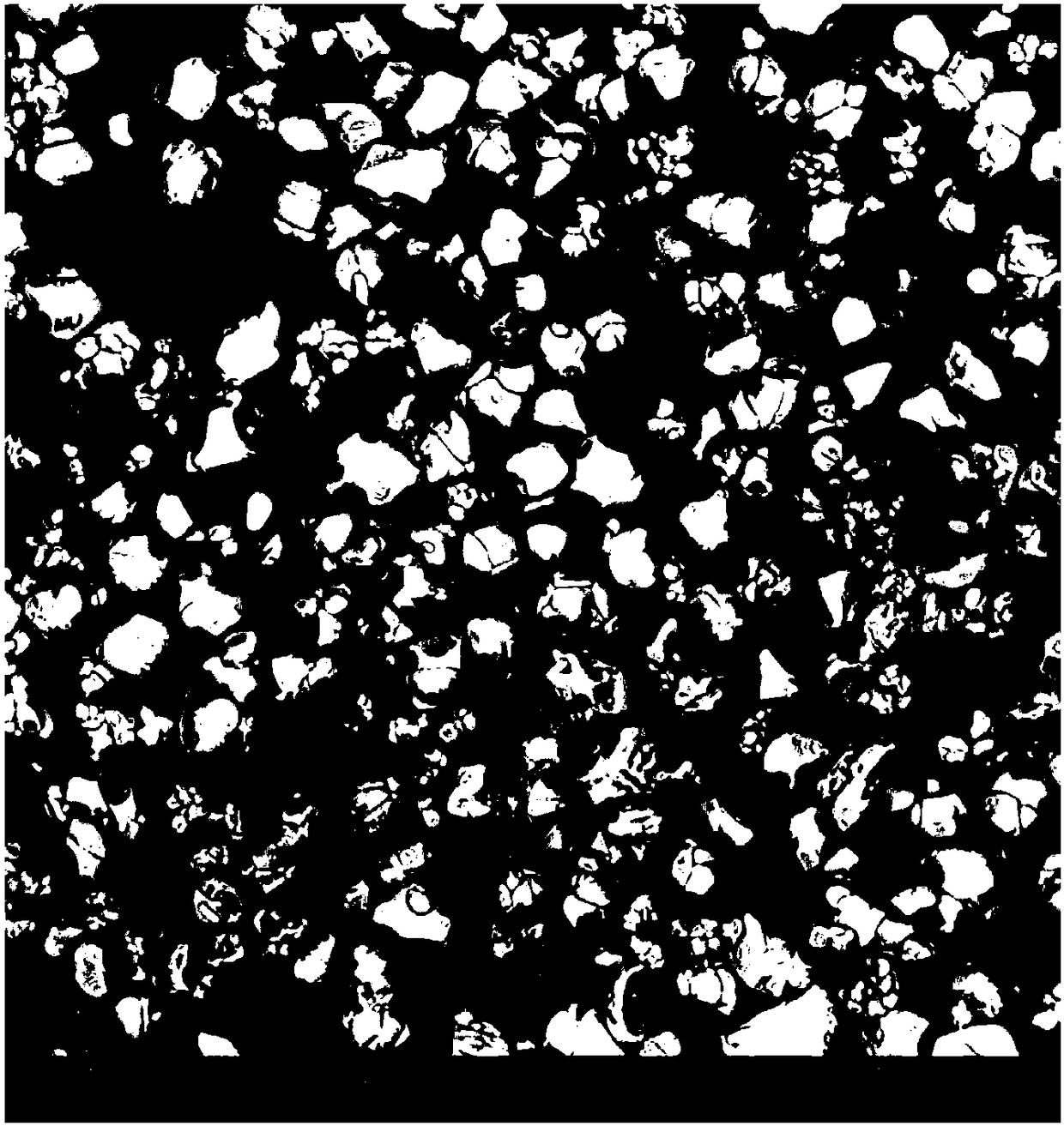
A method for preparing a micron ionic crystal powder sample for scanning electron microscopy
A technology of ion crystal and scanning electron microscope, which is applied in the direction of material analysis, measuring devices, and instruments using wave/particle radiation, can solve problems such as agglomeration and agglomeration, and achieve short sample preparation cycle, tight combination, and uniform powder dispersion Effect
- Summary
- Abstract
- Description
- Claims
- Application Information
AI Technical Summary
Problems solved by technology
Method used
Image
Examples
Embodiment 1
[0033] A method for preparing a scanning electron microscope sample of a micron-sized ionic crystal powder, comprising the steps of:
[0034] (1) Dispersion: Take the powder sample and the dispersion liquid according to a certain proportion, add the powder sample into the dispersion liquid and stir to disperse to obtain a suspension;
[0035] (2) Sampling: Take 1-2 drops of the suspension in step (1) in the mold and volatilize at a certain temperature. After the dispersion is volatilized, the dispersed powder sample is obtained;
[0036] (3) Prepare the resin glue: blend the resin and the curing agent in a certain proportion, and stir to obtain the resin glue for curing the sample;
[0037] (4) Cold mounting sample preparation: Pour the resin glue prepared in step (3) into the mold of step (2), and let it stand at a certain temperature until the sample is solidified to obtain a metallographic sample;
[0038] (5) Gold spraying: Sputtering gold spraying on the surface of the s...
Embodiment 2
[0048] A method for preparing a scanning electron microscope sample of a micron-sized ionic crystal powder, comprising the steps of:
[0049] (1) Dispersion: Take the powder sample and the dispersion liquid according to a certain proportion, add the powder sample into the dispersion liquid and stir to disperse to obtain a suspension;
[0050] (2) Sampling: Take 1-2 drops of the suspension in step (1) in the mold and volatilize at a certain temperature. After the dispersion is volatilized, the dispersed powder sample is obtained;
[0051] (3) Prepare the resin glue: blend the resin and the curing agent in a certain proportion, and stir to obtain the resin glue for curing the sample;
[0052] (4) Cold mounting sample preparation: Pour the resin glue prepared in step (3) into the mold of step (2), and let it stand at a certain temperature until the sample is solidified to obtain a metallographic sample;
[0053] (5) Gold spraying: Sputtering gold spraying on the surface of the s...
Embodiment 3
[0062] A method for preparing a scanning electron microscope sample of a micron-sized ionic crystal powder, comprising the steps of:
[0063] (1) Dispersion: Take the powder sample and the dispersion liquid according to a certain proportion, add the powder sample into the dispersion liquid and stir to disperse to obtain a suspension;
[0064] (2) Sampling: Take 1-2 drops of the suspension in step (1) in the mold and volatilize at a certain temperature. After the dispersion is volatilized, the dispersed powder sample is obtained;
[0065] (3) Prepare the resin glue: blend the resin and the curing agent in a certain proportion, and stir to obtain the resin glue for curing the sample;
[0066] (4) Cold mounting sample preparation: Pour the resin glue prepared in step (3) into the mold of step (2), and let it stand at a certain temperature until the sample is solidified to obtain a metallographic sample;
[0067] (5) Gold spraying: Sputtering gold spraying on the surface of the s...
PUM
| Property | Measurement | Unit |
|---|---|---|
| Particle size | aaaaa | aaaaa |
Abstract
Description
Claims
Application Information
 - R&D
- Intellectual Property
- Life Sciences
- Materials
- Tech Scout
- Unparalleled Data Quality
- Higher Quality Content
- 60% Fewer Hallucinations
Browse by: Latest US Patents, China's latest patents, Technical Efficacy Thesaurus, Application Domain, Technology Topic, Popular Technical Reports.
© 2025 PatSnap. All rights reserved.Legal|Privacy policy|Modern Slavery Act Transparency Statement|Sitemap|About US| Contact US: help@patsnap.com

