Method for automatically extracting tracheal tree from chest CT image

A CT image and tracheal tree technology, applied in the field of image processing based on medical images, can solve problems such as difficulty in completing CT image segmentation tasks, restricting the accuracy of tracheal segmentation, and missing detailed information

Active Publication Date: 2018-06-15
NORTHEASTERN UNIV
View PDF11 Cites 24 Cited by
  • Summary
  • Abstract
  • Description
  • Claims
  • Application Information

AI Technical Summary

Problems solved by technology

[0004] Due to the special topological structure and gray texture features of the pulmonary tracheal tree, as the tracheal tree divides step by step, the lumen becomes thinner and the tube wall becomes thinner, and the gray value of the tracheal wall gradually decreases on the CT image. , some segmentation methods routinely used for lung parenchyma, liver, and brain are not well suited for the segmentation of lung and trachea
Tracheal segmentation based on traditional threshold growth tends to spread across the tracheal wall into the lung parenchyma, forming a large-scale over-segmentation, that is, the "leakage" phenomenon, and it is difficult to effectively identify subtle or diseased branches, leaving a lot of detailed information, which is difficult to obtain accurate pulmonary tracheal tree
[0005] Among the currently disclosed tracheal tree processing methods, patent CN201210423958.2 uses multi-scale gray scale reconstruction to enhance the lumen area; on this basis, patent CN201510009239.X uses multi-scale tubular structure features to extract tracheal tree, both methods consume A lot of time; the patent CN201110405950.9 uses a fixed threshold to extract the bronchi below the segment level, and the designed leakage processing model is only for the segmental branches, it is difficult to obtain deeper bronchi and fine trachea, and there is no specific leakage processing method designed for these branches; Patent CN201510224781.7 utilizes energy function to reconstruct subtle and peripheral trachea, but still needs a fixed threshold to judge trachea membership and leakage
The above methods require strict manual parameter setting, or require complex preprocessing and enhancement operations on CT images, or need to extract tubular structural features and other auxiliary tracheal segmentation, which greatly increases the processing time and restricts the accuracy of tracheal segmentation, making it difficult to complete multiple The segmentation task of CT images under various imaging conditions seriously restricts the reliability and applicability in clinical applications

Method used


Image

  • Method for automatically extracting tracheal tree from chest CT image
  • Method for automatically extracting tracheal tree from chest CT image
  • Method for automatically extracting tracheal tree from chest CT image

Examples


Embodiment 1

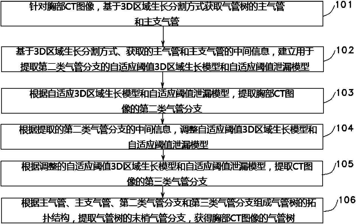
[0122] The technical problem solved by this embodiment is to provide a method for automatically extracting a tracheal tree from a chest CT image, using an adaptive threshold to avoid manual interaction and parameter setting required by the prior art, so as to adapt to various imaging conditions and pathological conditions tracheal tree segmentation task. like figure 1 As shown, the present invention provides a method for fully automatic extraction of trachea from chest CT images, and the technical solution is as follows:

[0123] 101. For the chest CT image, obtain the first type of tracheal branches of the tracheal tree based on the 3D region growth segmentation method, that is, the main trachea and the main bronchus, wherein the main bronchus includes the left main bronchus and the right main bronchus;

[0124] 102. Based on the 3D region growth segmentation method, the acquired intermediate information of the main trachea, and the acquired intermediate information of the m...

Embodiment 2

[0130] The main trachea and main bronchus are surrounded by a relatively complete and bright tracheal wall and separated from the lung parenchyma. The main trachea and main bronchus are extracted from the CT image without considering the leakage situation, which is less difficult. Therefore, the common threshold 3D region growth segmentation method is used. Effective access to the main trachea and main bronchi.

[0131] In this embodiment, the first type of tracheal branch is obtained from the chest CT image, including:

[0132] 1. Obtain the main trachea from chest CT images:

[0133] 1011. Read in the chest CT image, in order to avoid the influence of CT image noise on subsequent growth segmentation, perform a Gaussian smoothing preprocessing operation on the chest CT image with a three-dimensional scale of σ=0.5mm;

[0134] 1012. Obtain a layer of CT images of the preprocessed chest CT image from the chest top to the chest base, and perform image binarization processing on t...

Embodiment 3

[0152] In this embodiment, an adaptive threshold 3D region growth model and an adaptive threshold leakage model are established according to the acquired intermediate information such as trachea gray distribution, spatial scale, and segmentation process information of the main trachea and main bronchus.

[0153] The specific steps for establishing an adaptive threshold 3D region growth model are as follows:

[0154] 1021: Obtain an initial segmentation seed point set; the initial segmentation seed point set includes: all seed points in the segmentation queue of the left main bronchus when the iteration is terminated and all seed points in the segmentation queue of the right main bronchus when the iteration is terminated.

[0155] 1022: Obtain a grayscale threshold; in this embodiment, a certain grayscale threshold is set for the segmentation of the trachea tree to determine the upper limit of the trachea segmentation, and at the same time, the pixels whose grayscale values ​​ex...


PUM


Abstract

The invention belongs to the technical field of medical image-based image processing, and particularly relates to a method for automatically extracting a tracheal tree from a chest CT image. The method comprises the steps of obtaining main tracheae and main bronchi; according to a 3D region growing segmentation mode and information of the obtained main tracheae and main bronchi, building an adaptive threshold 3D region growing segmentation model and an adaptive threshold leakage model; by utilizing the adaptive threshold 3D region growing segmentation model and the adaptive threshold leakage model, extracting second tracheal branches of the chest CT image; according to intermediate information of the extracted second tracheal branches, adjusting parameters of the adaptive threshold 3D region growing segmentation model and the adaptive threshold leakage model, and then extracting third tracheal branches of the chest CT image; and based on an obtained tracheal tree topology structure, extracting terminal tracheal branches, and obtaining the tracheal tree of the chest CT image. According to the method provided by the invention, the tracheal segmentation precision of extracting the tracheal tree from the CT image is improved and the extraction time is shortened.

Description

technical field [0001] The invention belongs to the technical field of image processing based on medical images, and in particular relates to a method for automatically extracting a trachea tree from a chest CT image. Background technique [0002] Obtaining accurate pulmonary tracheal tree structure from CT images is of great significance in the medical and computer application world. Clinicians can conduct pathological analysis and follow-up research on common respiratory diseases such as chronic obstructive pulmonary disease and bronchiectasis through tracheal parameters and grading information; they can also perform non-invasive virtual bronchoscopy on patients; The anatomical one-to-one correspondence of sub-level lung structures such as lobes and segments is also an important basis for image segmentation and analysis of related structures. Therefore, lung trachea segmentation based on CT images has always been a hot spot of researchers. [0003] In the chest CT image,...

Claims


Application Information

Patent Timeline
Patent Type & Authority Applications(China)
IPC IPC(8): G06T7/00G06T7/11G06T7/136G06T7/187G06T7/194
CPCG06T7/0012G06T7/11G06T7/136G06T7/187G06T7/194G06T2207/10081G06T2207/20156G06T2207/30061
Inventor 边子健覃文军杨金柱栗伟曹鹏冯朝路魏星王同亮林国丛刘欢迎杨琦赵大哲
Owner NORTHEASTERN UNIV
Why Eureka
Try Eureka