
Microexamination method for forms of fishes laying drifting eggs at different developmental stages of early phase

A developmental stage, microscopic observation technology, applied in fish farming, application, climate change adaptation, etc., can solve the problems of difficult to freeze pictures to capture developmental details, difficult to obtain complete embryo images, and easy to move live larvae. Complete, vivid shooting effect, less bleaching effect

Active Publication Date: 2018-10-16
JIANGXI INST OF AQUATIC SCI
View PDF8 Cites 0 Cited by
  • Summary
  • Abstract
  • Description
  • Claims
  • Application Information

AI Technical Summary

Problems solved by technology

[0004] The present invention aims at the double flow caused by floating fish roe in the early development process due to small embryos and too large egg membranes, fertilized eggs suspended in water, and embryos suspended in egg membranes, and it is difficult to find the best one. Shooting angle; secondly, after the embryo develops to the muscle effect stage, the embryo shrinks in the egg membrane, and it is difficult to obtain a complete embryo image; live larvae are very prone to hyperactivity, and it is difficult to freeze the picture to capture development details. Microscopic observation method of morphology in different early developmental stages of genus

Method used


Image

  • Microexamination method for forms of fishes laying drifting eggs at different developmental stages of early phase
  • Microexamination method for forms of fishes laying drifting eggs at different developmental stages of early phase
  • Microexamination method for forms of fishes laying drifting eggs at different developmental stages of early phase

Examples


Embodiment 1

[0041] A microscopic observation method for the early stages of different developmental stages of fishes that lay drifting eggs. Taking the eggs of goblins in the blastopore closed stage as an example, the observation method is as follows:

[0042] (1) Use a straw or a small spoon to take a goblin egg from the blastopore closed stage, and put it in the central area of ​​a clean and dry petri dish;

[0043] (2) Use a dry small strip-shaped sponge to absorb the moisture between the fish eggs and the petri dish, so that the fish eggs can be firmly placed on the petri dish;

[0044] (3) Place the above-mentioned Petri dish with fish eggs under the objective lens of the dissecting mirror, and use a dry small strip sponge to move the fish eggs to the desired viewing or shooting angle;

[0045] (4) Adjust the dissecting mirror, obtain the observation or photographing images, and complete the observation or photographing within 5 minutes;

[0046] The port area of ​​the small elongat...

Embodiment 2

[0048] A microscopic observation method for the morphology of fishes that lay drifting eggs at different early stages of development, taking the eggs of goblins in the heartbeat stage as an example, the observation method is as follows:

[0049] (1) Use a straw or a small spoon to take a piece of goblin roe during the heartbeat period, and put it in the central area of ​​a clean and dry petri dish;

[0050] (2) Use a dry small strip-shaped sponge to absorb the water between the fish eggs and the petri dish, so that the fish eggs can be firmly placed on the petri dish; the port area of ​​the small strip-shaped sponge does not exceed 0.1cm 2 .

[0051](3) Place the above-mentioned petri dish with fish eggs under the objective lens of the dissecting mirror, adjust the microscopic magnification of the dissecting mirror, and use a thread needle to pierce the fish egg membrane from the left and right sides of the fish eggs, and the vertical height of the puncture point is Fish roe ...

Embodiment 3

[0058] A kind of microscopic observation method of different early stages of development of fishes that lay drifting eggs, taking mandarin fish as an example, the observation method is as follows:

[0059] (1) Take a mandarin fish with a straw or a small spoon, and put it in the central area of ​​a clean and dry petri dish;

[0060] (2) Use a 10ml syringe to add or absorb the water in the contact part of the larvae and the petri dish to 0.5ml;

[0061] (3) Use a 10ml syringe to drop 0.08ml of 90% ethanol around the larvae, let it stand for 2 minutes, then use a 10ml syringe to drop 0.08ml of 50% ethanol around the larvae, and let it stand for 3 minutes;

[0062] (4) Control 0.35ml liquid to cover the larvae; the control method is: 10ml syringe absorbs excess liquid;

[0063] (5) Move the petri dish until the larvae are within the observation field of the dissecting mirror, adjust the multiple of the dissecting mirror, obtain the observation or photographing picture, and compl...


PUM


Abstract

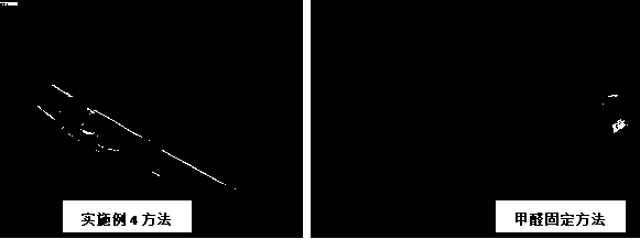
The invention discloses a microexamination method for the forms of fishes laying drifting eggs at different developmental stages of the early phase. The microexamination method is characterized by being composed of a development form observation method from fertilization of the drifting eggs to the tail bud stage, a development form observation method from the muscle effect stage to the early hatching stage and a development form observation method of hatched fry. According to the method, the problems that due to the fact that from fertilization to the tail bud stage of the drifting eggs, theembryos are small, and the egg envelopes are large, the germ cells suspend in water, the embryos suspend in the egg envelopes and flow in pairs, and the best shooting angle is hard to find can be solved. Then the problems that from the muscle effect stage to the early hatching stage, the embryos crouch in the egg envelopes and turn over, and a complete embryo image is hard to obtain are solved. Finally, the problem that due to the fact that the fry is hyperactive easily after being hatched, the shooting picture cannot be fixed is solved. Practice proves that the method is used for microexamination of the drifting eggs or the fry, the shooting angle is adjustable, the embryo development picture is easy to capture, the fry rarely fades or dehydrates, the shooting effect is alive, and the details are complete.

Description

technical field [0001] The invention relates to a microscopic observation method for the morphology of different early developmental stages of floating egg-laying fish, which belongs to the field of fish morphology observation. Background technique [0002] Fishes that lay drifting eggs are widely distributed in inland river systems. Live photography of their eggs and larvae is an important means of studying the early life history of fishes. It is currently a common method to observe the morphological development of fish eggs and larvae by fixing them with a fixative solution. This method has the problems of poor observation and photographing effects, and the need to change the fixative solution frequently. The steps are complicated and easy to store and take out. Destruction of specimen structure, strong pungent smell of fixative, etc. The fertilized eggs of floating fish eggs are transparent, and the embryonic development can be directly observed under the dissecting micr...

Claims


Application Information

Patent Timeline
Patent Type & Authority Applications(China)
IPC IPC(8): A01K61/10A01K61/17
CPCA01K61/10A01K61/17Y02A40/81
Inventor 王生傅义龙章海鑫饶毅徐先栋张爱芳张燕萍李艳芳张桂芳万锦涛
Owner JIANGXI INST OF AQUATIC SCI
Learn More