Single-scanning quantitative magnetic resonance T2 * imaging method based on residual network reconstruction

A technology of magnetic resonance imaging and imaging method, which is applied in the directions of using nuclear magnetic resonance imaging system for measurement, magnetic resonance measurement, and magnetic variable measurement, etc. It can solve the problem of difficulty in separating four overlapping echo signals, and achieve a large measurement range. Effect

Active Publication Date: 2018-10-16
XIAMEN UNIV
View PDF6 Cites 10 Cited by
  • Summary
  • Abstract
  • Description
  • Claims
  • Application Information

AI Technical Summary

Problems solved by technology

However, it is difficult to separate the 4 overlapping echo signals by conventional methods

Method used


Image

  • Single-scanning quantitative magnetic resonance T2 * imaging method based on residual network reconstruction
  • Single-scanning quantitative magnetic resonance T2 * imaging method based on residual network reconstruction
  • Single-scanning quantitative magnetic resonance T2 * imaging method based on residual network reconstruction

Examples


specific Embodiment

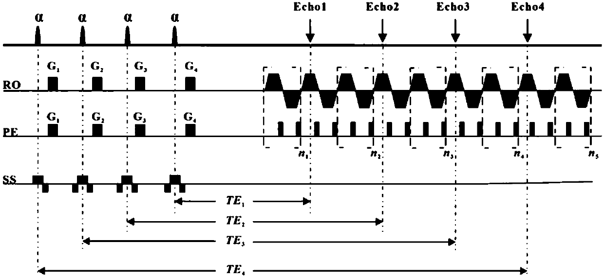
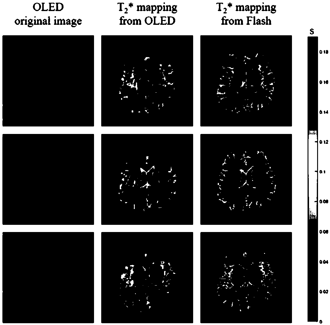
[0066] Single-scan quantitative magnetic resonance T based on residual network reconstruction algorithm 2 *The quantitative imaging method has been carried out in human brain experiments to verify the feasibility of the present invention. The experiment was carried out under the human MRI 3T imager. On the operating table of the magnetic resonance imager, open the corresponding operating software in the imager, first locate the region of interest for the imaging object, and then perform tuning, shimming, power and frequency correction. In order to evaluate the effectiveness of the image obtained by this method, a Flash imaging experiment was carried out in the same environment as a comparison. Then import the compiled OLED imaging sequence (such as figure 1 (Shown), according to specific experimental conditions, set the various parameters of the pulse sequence, the experimental parameters of this embodiment are set as follows: the imaging field FOV is 22cm×22cm, the excitation ...


PUM


Abstract

A single-scanning quantitative magnetic resonance T2 * imaging method based on residual network reconstruction relates to a magnetic resonance imaging method. Four small-angle excitation pulses with the same deflection angle are utilized, a period of evolution time is formed after each excitation pulse, so that the transverse relaxation time of each echo signal is different. A shifting gradient ofa frequency dimension and a phase dimension is added after each excitation pulse, so that the signals generated by different excitation pulses are different in the position of the k space. In this way, a plurality of gradient echo signals with different transverse relaxation time are obtained in one-time sampling. The sampling signals are subjected to normalization, zero filling and fast fouriertransformation and then input to a trained residual network to be reconstructed to obtain a quantitative T2 * image. The training data of the residual network are derived from simulation data, and theinput image of a network is obtained via a random generation template through simulating the experiment environment for sampling. The template serves as a label, and a mapping relation between an input image and an output image is obtained through training.

Description

Technical field [0001] The invention relates to a magnetic resonance imaging method, in particular to a single-scan quantitative magnetic resonance imaging method based on a residual network reconstruction algorithm. 2 *Imaging method. Background technique [0002] Magnetic resonance parametric imaging (for example, T 1 Imaging, T 2 Imaging and T 2 * Imaging) can be easily used to provide quantitative information for characterizing specific tissue properties [1] . Quantitative imaging has many outstanding features, among which the most obvious advantage is that it can eliminate the influences that are independent of the nature of the tissue, such as dependence on the operator, differences in scanning parameters, spatial changes in magnetic fields and image scaling, etc. [2] . T 2 *Imaging, as a kind of MRI quantitative imaging, provides a contrast mechanism for analyzing normal and diseased living tissues in a non-invasive way. 2 *Relaxation measurement can get the iron content o...

Claims


Application Information

Patent Timeline
Patent Type & Authority Applications(China)
IPC IPC(8): G01R33/48G01R33/56
CPCG01R33/48G01R33/56
Inventor 蔡聪波王超丁兴号蔡淑惠陈忠
Owner XIAMEN UNIV
Why Eureka
Try Eureka