Dual-mode probe combined with ultrasonic imaging and optical coherence tomography imaging

An optical coherence tomography and ultrasonic technology, applied in the field of medical devices, can solve the problems of complex assembly, only one to two millimeters, and technical difficulty, so as to ensure stability and reliability, ensure product consistency, and reduce process complexity. degree of effect

Pending Publication Date: 2018-11-13
INNERMEDICAL CO LTD
View PDF0 Cites 0 Cited by
  • Summary
  • Abstract
  • Description
  • Claims
  • Application Information

AI Technical Summary

Problems solved by technology

However, because OCT uses near-infrared light as a detection method, its frequency is much higher than that of ultrasonic waves. Therefore, the resolution of OCT images (a few microns to a dozen microns) is more than 10 times that of ultrasonic images. The disadvantage is that the imaging depth of OCT is only ( One to two millimeters), so the imaging depth of the two is very different
In recent years, although universities and research institutions have been developing dual-mode imaging probes that combine ultrasound and OCT, due to technical difficulties, complex processes, complex assembly, and differences in imaging depth, the stability and reliability of the work are still improving. Therefore, there is no such dual-mode probe available in clinical practice at present.

Method used


Image

  • Dual-mode probe combined with ultrasonic imaging and optical coherence tomography imaging
  • Dual-mode probe combined with ultrasonic imaging and optical coherence tomography imaging
  • Dual-mode probe combined with ultrasonic imaging and optical coherence tomography imaging

Examples


Embodiment 1

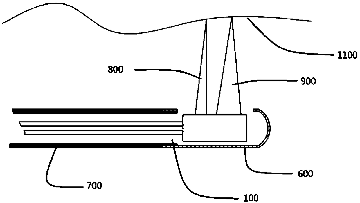
[0053] like Figure 1~Figure 2 As shown, a dual-mode probe combining ultrasonic imaging and optical coherence tomography includes a torque transmission component 700 , a protective cover 600 and a dual-mode imaging core 100 . The protective sleeve 600 is rigidly connected with the torque transmission component 700 away from the joint end, and is used to protect the imaging components of the dual-mode imaging core 100; the dual-mode imaging core 100 is located inside the catheter core composed of the torque transmission component 700 and the protective sleeve 600, The near-infrared beam 800 and the ultrasonic beam 900 are emitted from the same side of the dual-mode imaging core 100 , and reach the biological tissue 1100 through the exit window of the protective cover 600 .

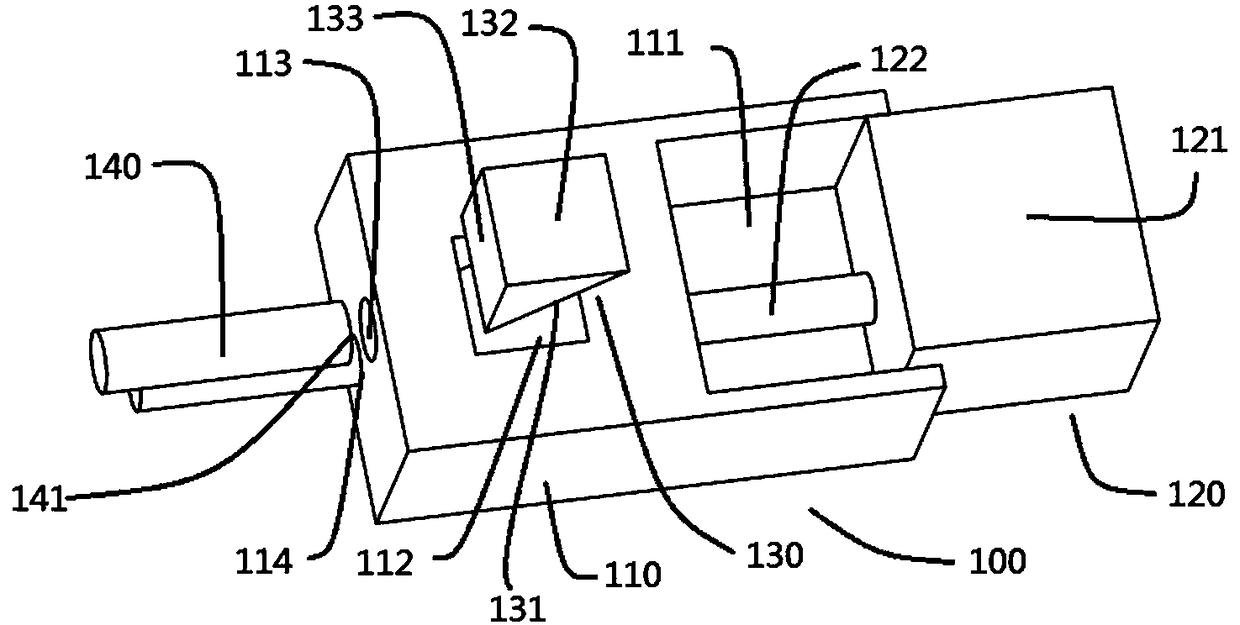
[0054] The dual-mode imaging core 100 can have three different structures. This example figure 2 As shown, the dual-mode imaging core 100 includes a base 110 and imaging components, and the imaging compo...

Embodiment 2

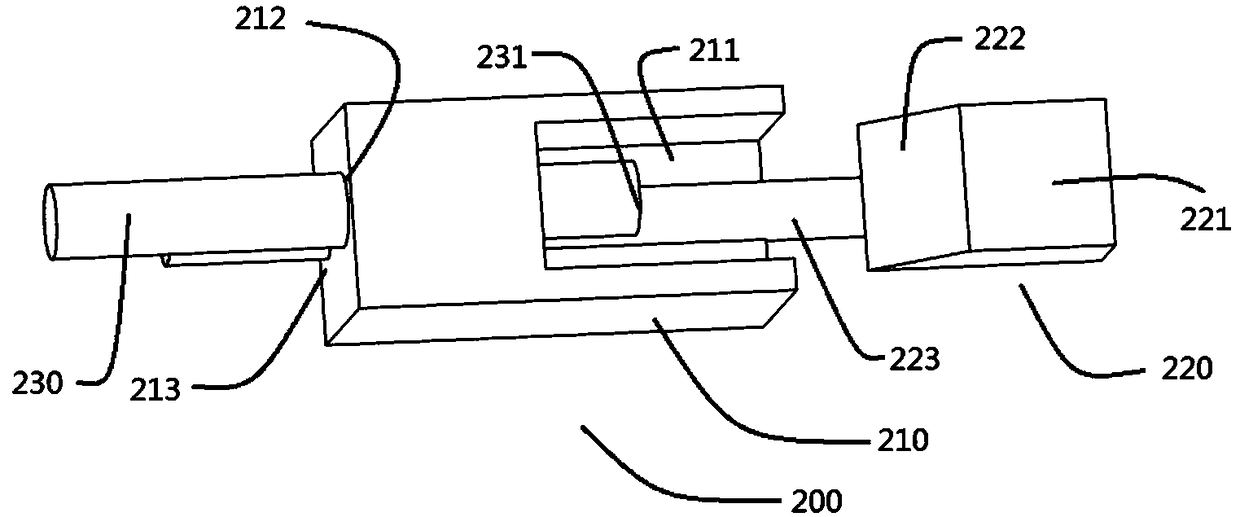
[0057] On the basis of Example 1, such as figure 1 and image 3 As shown, the dual-mode imaging core 200 of this example is based on the base 210, the ultrasonic transducer 220 is rigidly fixed on the ultrasonic transducer installation groove 211 of the base 210, and the coaxial cable 223 of the ultrasonic transducer 220 Pass through the coaxial cable installation hole 213 of the base 210 and fix it. In this example, if image 3 As shown, the beam turning surface 222 of the beam turning component is arranged on the rear end surface of the ultrasonic transducer 220, the light guiding element and the focusing element 230 are fixedly installed in the light guiding element mounting hole 212, and the infrared light is transmitted by the light guiding element and the focusing element 230. The focusing element beam exit surface 231 of the focusing element 230 emits, irradiates on the beam turning surface 222 of the rear end surface of the ultrasonic transducer 220, and turns it int...

Embodiment 3

[0059] On the basis of Example 1, such as figure 1 and Figure 4 As shown, the dual-mode imaging core 500 is based on the base 510, the ultrasonic transducer 520 is rigidly fixed on the ultrasonic transducer installation groove 511 of the base 510, and the coaxial cable 522 of the ultrasonic transducer 520 is connected from the base Pass through the coaxial cable installation hole 515 of the seat 510 and fix it. A groove 512 is provided on the base 510 , and the beam refracting surface 513 of the beam deflection component is arranged adjacent to the groove 512 and at one end close to the ultrasonic transducer installation groove 511 . The light guide element and the focus element 530 are fixedly installed in the light guide element installation hole 514, the infrared light is emitted from the focus element beam exit surface 531 of the light guide element and focus element 530, and the beam irradiated on the base 510 is deflected surface 513 , and turn to emit in a direction ...


PUM


Abstract

The invention provides a dual-mode probe combined with ultrasonic imaging and optical coherence tomography imaging, which comprises a dual-mode imaging core module, wherein the dual-mode imaging coremodule comprises a base and an imaging component, the imaging component comprises a light guide element and a focusing element, a ligh beam transition component and a ultrasonic transducer, the ultrasonic transducer, the light guide element and the focusing element are respectively fixed on a base, the light beam transition component is a independent element which is fixedly arranged on the base or a light beam transition surface arranged on the base or on the back side surface of the ultrasonic transducer, the light beam emerging surfaces of the light guide element and focusing element face towards the light beam transition component; light is emitted from the light guide element and the focusing element and passes through the light beam transition component, a ultrasonic beam is emittedfrom the ultrasonic transducer, and the direction of the light emerging is the same as or opposite to the direction of the ultrasonic beam emerging. According to the technical scheme, the base is utilized to fix the ultrasonic and optical elements, the stability and reliability of working are guaranteed, and the ultrasound and OCT images of diseased tissues can be collected at one time.

Description

technical field [0001] The invention belongs to the technical field of medical devices, in particular to a dual-mode probe combined with ultrasonic imaging and optical coherence tomography. Background technique [0002] At present, ultrasonic imaging technology and OCT (optical coherence tomography) technology are relatively mature in the field of medical endoscopic imaging, and are widely used in clinical disease diagnosis and surgical assistance. In endoscopic imaging technology, the probe is the core component of the endoscopic imaging system, which directly determines the image quality. Ultrasound has better penetrability to biological tissues, and can reach tens of millimeters to several centimeters, and the imaging depth is much greater than that of OCT. However, because OCT uses near-infrared light as a detection method, its frequency is much higher than that of ultrasonic waves. Therefore, the resolution of OCT images (a few microns to a dozen microns) is more than ...

Claims


Application Information

Patent Timeline
Patent Type & Authority Applications(China)
IPC IPC(8): A61B8/00A61B5/00
CPCA61B8/4444A61B5/0066
Inventor 白晓淞黄赞力
Owner INNERMEDICAL CO LTD
Why Eureka
Try Eureka