Fusion method for coronary artery CT image and cardiac ultrasonic strain image

A technique of coronary artery and CT imaging, which is applied in the field of biomedical engineering, can solve the problems of evaluating the degree of myocardial ischemia, cost, and comprehensive evaluation of the disease state.

Active Publication Date: 2018-11-13
BEIJING ANZHEN HOSPITAL AFFILIATED TO CAPITAL MEDICAL UNIV
View PDF7 Cites 18 Cited by
  • Summary
  • Abstract
  • Description
  • Claims
  • Application Information

AI Technical Summary

Problems solved by technology

However, the test is radioactive, expensive and time-consuming
Therefore, it is limited to assess the extent of myocardial ischemia in severe and young patients.
[0005] (3) Conventional echocardiography (UCG) mainly reflects the overall and local systolic function of the left ventricle by measuring the left ventricular ejection fraction and wall motion thickening rate, but the sensitivity is low, and it is difficult to detect myocardial damage and dysfunction early
[0006] Although single modality imaging technologies have their own strengths, they cannot achieve multi-level and comprehensive assessment of disease status

Method used


Image

  • Fusion method for coronary artery CT image and cardiac ultrasonic strain image
  • Fusion method for coronary artery CT image and cardiac ultrasonic strain image
  • Fusion method for coronary artery CT image and cardiac ultrasonic strain image

Examples


Embodiment 1

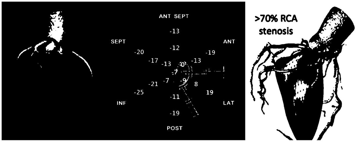
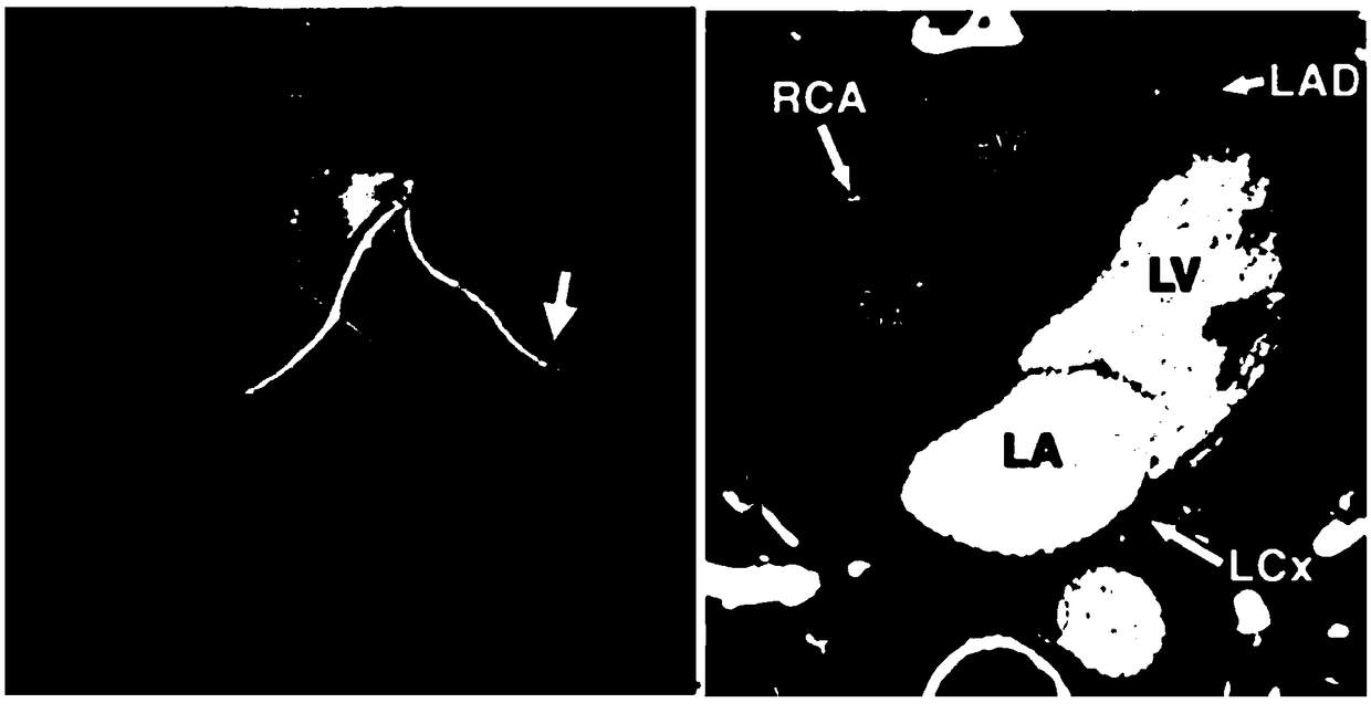
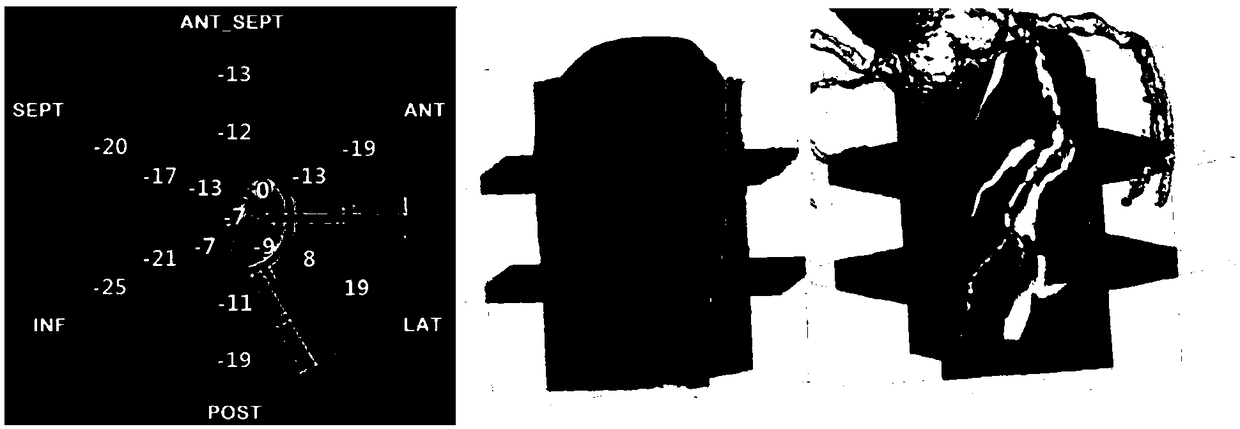
[0020] 1. The positioning method of "one point, one side and three planes" performs three-dimensional fusion of cardiac ultrasound images and coronary artery CT images.

[0021] The present invention fuses cardiac ultrasound images and CT images for the first time, and fuses echocardiograms and coronary artery CT images through a positioning method of "one point, one plane and three planes", and displays a three-dimensional model diagram.

[0022] Image fusion is a continuous process, and the basic steps include: (1) Image acquisition and import: Coronary artery CT examination in patients with coronary heart disease, obtaining the original data in DICOM format of coronary artery CT in patients with coronary heart disease. Patients underwent echocardiography, image analysis was performed using ultrasound strain techniques, and data were stored in raw DICOM format. (2) Image registration and fusion: Since there are huge differences between echocardiographic images and CT images ...


PUM


Abstract

The invention relates to a fusion method for a coronary artery CT image and a cardiac ultrasonic strain image. The invention provides a multi-mode image fusion technology method for fusing and mappinga two-dimensional UCG image and a three-dimensional CT image to stereoscopically display coronary artery lesions and myocardial damage conditions. According to the method, The cardiac ultrasonic image and the CT image are fused for the first time; the ultrasonic cardiogram and the coronary artery CT image are fused (figure 1) through a one-point one-surface three-section positioning method; and athree-dimensional pattern diagram is displayed. Then, on the basis of a three-dimensional ultrasonic volume image, two-dimensional images (figure 2) of 20 sections and 480 grid segments of a left ventricular myocardium are obtained by adopting a central axis rotation method and a refined segmentation method; based on this, the strain values of 480 segments are obtained; and myocardial perfusion ischemia information obtained by three-dimensional strain is superimposed into the three-dimensional coronary artery vascular pattern diagram (figure 3), thereby accurately reflecting a relation between a myocardial ischemia area and an actual coronary artery stenosis.

Description

technical field [0001] A fusion method of coronary artery CT images and cardiac ultrasound (UCG) strain imaging proposed by the present invention belongs to the technical field of biomedical engineering, and relates to a multimodal image fusion for human coronary artery CT images and cardiac ultrasound strain imaging method. Background technique [0002] With the rapid development of imaging technology, imaging assessment techniques for coronary heart disease have become more diverse and mature. Commonly used imaging methods include CT, SPECT, and UCG. [0003] (1) Coronary artery CT is the "gold standard" for evaluating coronary artery stenosis. Although coronary CT can provide information similar to coronary angiography, such as structural abnormalities such as stenosis, it does not necessarily correlate well with hemodynamic status. Coronary CT cannot determine the true hemodynamic state of the stenosis and myocardial ischemia perfusion, and CT is an invasive examinati...

Claims


Application Information

Patent Timeline
Patent Type & Authority Applications(China)
IPC IPC(8): G06T7/30
CPCG06T2207/10081G06T2207/10136G06T2207/20221G06T2207/30048G06T7/30
Inventor 杨娅谢谨捷乔爱科
Owner BEIJING ANZHEN HOSPITAL AFFILIATED TO CAPITAL MEDICAL UNIV
Why Eureka
Try Eureka