Positioning method of CT image for PET attenuation correction

A CT image and positioning method technology, applied in the field of medical devices, can solve the problems of insufficient statistics of transmission scan data, differences, and influence on the accuracy of attenuation correction.

Active Publication Date: 2018-11-20
RAYSOLUTION DIGITAL MEDICAL IMAGING CO LTD
View PDF5 Cites 2 Cited by
  • Summary
  • Abstract
  • Description
  • Claims
  • Application Information

AI Technical Summary

Problems solved by technology

The advantage of this method is that both the transmission scan and the emission scan are 511keV gamma-ray scans, and the obtained attenuation coefficient does not need to be converted between photon energies; the disadvantage is that the transmission scan is easily polluted by the emission count in the subject; Due to the limitations of time and radiation source dose, the statistics of transmission scan data are often insufficient, which affects the accuracy of attenuation correction; due to the limitation of the g

Method used


Image

  • Positioning method of CT image for PET attenuation correction

Examples


Embodiment Construction

[0025] When performing a PET / CT examination, two sets of images must be acquired for a complete PET / CT scan, that is, one set for PET and one set for CT. The basic tissue structure image and the "attenuation image" required for attenuation correction are provided by CT, the attenuation correction is performed on the PET image, and the fusion of the PET image and the CT image is completed. The working process includes two parts: data acquisition and image processing and diagnosis, that is: firstly, patient data acquisition---that is, CT plain film positioning scan first, then CT scan, and then PET scan.

[0026] The specific scanning process is as follows: start PET / CT, first start CT and then PET, (when shutting down, turn off PET first, then CT), perform self-inspection of CT and PET, including tube warm-up and CT detector calibration. First, log in the patient, enter patient information such as patient name, age, injection of PET drug activity dose, etc. on the acquisition c...


PUM


Abstract

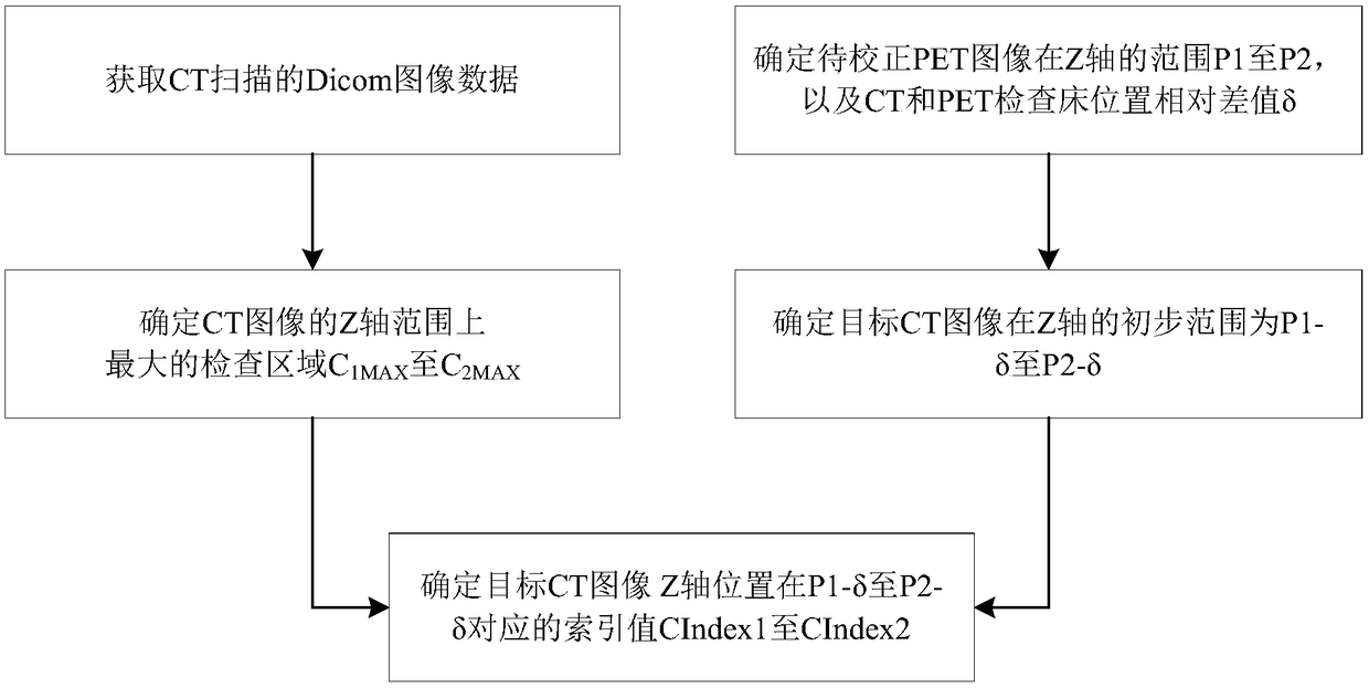
A positioning method of a CT image for PET attenuation correction comprises the following steps: (1) acquiring Dicom image data scanned by a CT, and determining a maximum inspection region C1MAX to C2MAX from the Dicom image data; 2) determining that a range of a PET image to be corrected in the Z-axis is P1 to P2, and determining a relative difference delta between positions of the CT and a PET examining table to determine that an initial value range of a target CT image in the Z-axis is P1-delta to P2-delta, (3) traversing the Dicom data of the CT image, determining index values CIndex1 to CIndex2 corresponding to the Z-axis position of the target CT image at the P1-delta to the P2-delta, wherein the CIndex1 is a maximum value index where the Z-axis position of the target CT image is less than a Z-axis minimum value of the PET image, the CIndex2 is a minimum value index where the Z-axis position of the target CT image is greater than the Z-axis minimum value of the PET image, and theCT image between the CIndex1 and the CIndex2 is a value range of the target CT image. The positioning method can effectively determine the number of layers of the target CT image corresponding to thePET image to be corrected, and is used for attenuation correction of the PET image to be corrected.

Description

technical field [0001] The invention belongs to the field of medical equipment, and relates to PET / CT imaging equipment, especially the image reconstruction technology of PET / CT imaging equipment. Background technique [0002] Positron emission tomography (PET) technology is an important part of modern medical imaging technology. Quantitative analysis of the metabolic process of tracers in the body is an important advantage of its imaging, but it has the greatest impact on the quantitative accuracy of PET images. A serious factor is the ray attenuation during the imaging process. Based on the characteristics of PET conforming to the imaging, the attenuation correction of the data can be completed by the method of transmission scanning. [0003] Early PET mostly used the method of 68Ge rod source transmission scanning for attenuation correction. In this method, an external 68Ge rod source is used to perform a standard blank scan first, and then a transmission scan is perform...

Claims


Application Information

Patent Timeline
IPC IPC(8): G06T11/00
CPCG06T11/003G06T11/005G06T2207/10081
Inventor 徐驰
Owner RAYSOLUTION DIGITAL MEDICAL IMAGING CO LTD
Why Eureka
Try Eureka