Tissue engineering bone blood vessel radiography method and blood vessel intubation tube

A tissue-engineered bone and angiography technology, used in medical science, instruments for radiological diagnosis, diagnosis, etc., can solve problems such as stable injection of unfavorable contrast agents, inability to study osteogenesis at the same time, blood incompatibility, etc. Conducive to stable injection, easy observation and easy operation

Pending Publication Date: 2018-12-07
STOMATOLOGY AFFILIATED STOMATOLOGY HOSPITAL OF GUANGZHOU MEDICAL UNIV
View PDF8 Cites 4 Cited by
  • Summary
  • Abstract
  • Description
  • Claims
  • Application Information

AI Technical Summary

Problems solved by technology

But it has obvious disadvantages. First, the operation is more complicated. Since Microfil is an oily solvent, it is incompatible with blood. Before angiography, it is necessary to wash the blood in the animal body with normal saline and inject formalin to fix the blood vessel. , the last is to inject the Microfil solvent
Furthermore, after using this method for angiography, the tissue-engineered bone must be decalcified before the vascularization can be detected on the computer. During the decalcification process, the new bone of the tissue-engineered bone will inevitably be decalcified, resulting in no imaging on CT. Therefore, this This method can only be used to study vascularization alone, and cannot study osteogenesis at the same time
[0003] Currently commercially available vascular cannula for experimental animals, during angiography, the cannula is easy to fall off, which is not conducive to the stable injection of contrast agent

Method used


Image

  • Tissue engineering bone blood vessel radiography method and blood vessel intubation tube
  • Tissue engineering bone blood vessel radiography method and blood vessel intubation tube
  • Tissue engineering bone blood vessel radiography method and blood vessel intubation tube

Examples


Embodiment Construction

[0018] The present invention will be further described below in conjunction with the examples. The description of the following examples is provided only to aid the understanding of the present invention. It should be pointed out that for those skilled in the art, without departing from the principles of the present invention, some improvements and modifications can be made to the present invention, and these improvements and modifications also fall within the protection scope of the claims of the present invention.

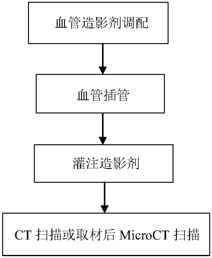
[0019] Taking the tissue engineering scaffold implanted in the experimental animal New Zealand rabbit as an example, this technology analyzes the vascularization effect of the tissue engineering scaffold in the animal body, and can also analyze the osteogenesis effect at the same time. The described tissue engineering bone angiography method comprises the following steps:

[0020] 1. Preferably adopt the ratio of 50-80% red lead oxide and 3-9% gelatin, and stir ...


PUM


Abstract

The invention relates to a tissue engineering bone blood vessel radiography method. The method includes the steps of firstly, blending a blood vessel radiography agent; secondly, conducting blood vessel tube intubation; thirdly, conducting blood vessel radiography; fourthly, putting an animal body in a refrigerator at 0-4 DEG C overnight, conducting CT scanning or taking a sample from a tissue engineering bone and putting the sample in formalin to be fixed for MicroCT scanning and analyzing. The method has the advantages that blood vessel casting agent lead oxide and a gelatin solution used inthe human anatomy field are adopted, a solution blended according to the ratio is directly injected into animal blood vessels, operation is simple, the bone formation situation and vascularization situation can be displayed on a CT three-dimensional image at the same time without decalcification, and the research on the relationship between bone formation and vascularization in the bone tissue engineering field is facilitated; a protruding structure is arranged in a blood vessel intubation tube, the disengagement of the intubation tube in the blood vessel radiography process is effectively prevented, and the stable injection of the radiography agent is facilitated.

Description

technical field [0001] The patent of the present invention relates to an angiography method, in particular to a tissue engineering bone angiography method and a blood vessel cannula. Background technique [0002] At present, in animal experiments in the field of tissue engineering, the contrast agent used to study vascularization is generally Microfil, which has a better effect on branch angiography, and can display the growth of blood vessels on machines such as MicroCT, which is beneficial for researchers to understand the shape and shape of blood vessels. Quantitative research on quantity, volume, density, etc. But it has obvious disadvantages. First, the operation is more complicated. Since Microfil is an oily solvent, it is incompatible with blood. Before angiography, it is necessary to wash the blood in the animal body with normal saline and inject formalin to fix the blood vessel. , the last is to inject the Microfil solvent. Furthermore, after adopting this method ...

Claims


Application Information

Patent Timeline
Patent Type & Authority Applications(China)
IPC IPC(8): A61B6/00
CPCA61B6/481A61B6/505
Inventor 周苗杨晓彬车月娟吴刚柳毅李树祎周卉周杨黄文燕李云阳
Owner STOMATOLOGY AFFILIATED STOMATOLOGY HOSPITAL OF GUANGZHOU MEDICAL UNIV
Why Eureka
Try Eureka