
Immunofluorescence kit for detecting human prostate cancer antigens PSMA and AR-V7 and application

A technology for prostate cancer and immunofluorescence detection, applied in biological testing, material inspection products, etc., can solve the problems of repeated sampling difficulties, a single tissue sample is not enough to reflect the tumor burden, and cannot be dynamically monitored

Inactive Publication Date: 2018-12-07
北京莱尔生物医药科技有限公司
View PDF10 Cites 0 Cited by
  • Summary
  • Abstract
  • Description
  • Claims
  • Application Information

AI Technical Summary

Problems solved by technology

[0010] At present, the pathological detection of tumor patients is mainly histological detection, which is faced with the difficulty of repeated sampling, cannot be dynamically monitored, and a single tissue sample is not enough to reflect the overall tumor burden, and cannot effectively reflect the non-humoral rare nuclei entering the body fluid in real time. cell changes

Method used


Image

  • Immunofluorescence kit for detecting human prostate cancer antigens PSMA and AR-V7 and application
  • Immunofluorescence kit for detecting human prostate cancer antigens PSMA and AR-V7 and application
  • Immunofluorescence kit for detecting human prostate cancer antigens PSMA and AR-V7 and application

Examples


Embodiment 1

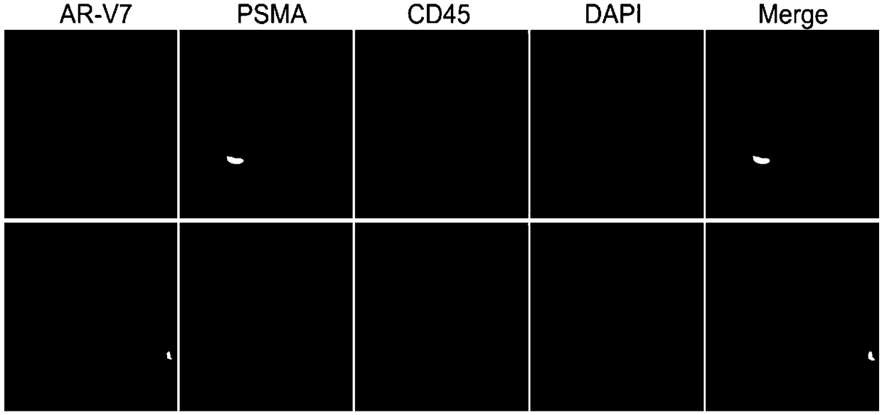
[0080] Materials: smears of negatively enriched blood samples, and 22RV1 and LNCaP cell smears for control cells.

[0081] Experimental steps:

[0082] 1. Draw 3.5ml of peripheral blood into an ACD (sodium citrate) anticoagulant tube. use The human peripheral blood leukocyte depletion kit negatively enriches tumor cells and fixes them on glass slides;

[0083] 2. Wash slides with CYP1 for 3 minutes x 3 times, 100-150 μL each time, to ensure that the entire sample area is covered;

[0084] 3. Absorb the excess liquid on the slide, add CYPP for 5 minutes, wash the slides with CYP1 as above for 3 minutes × 1 time; absorb excess liquid, add 200 μl of ice acetone:methanol (7:3) for 5 minutes, and wash the slides with CYP1 for 3 minutes × 3 times , to absorb excess water;

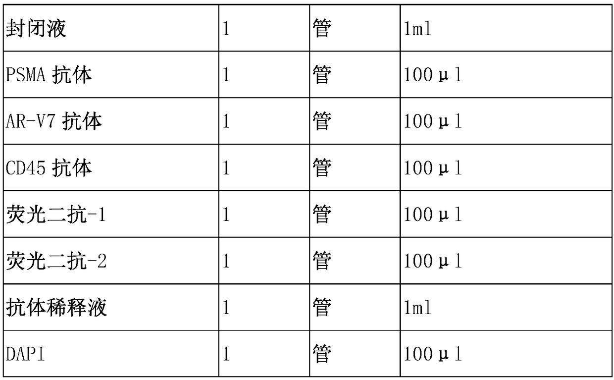
[0085] 4. Add 100-150 μl of blocking solution to block at room temperature for 25-30 minutes. Absorb excess blocking solution, add 100 μl of diluted PSMA antibody, AR-V7 antibody and CD45 antibody, and incu...

Embodiment 2

[0095] Materials: 1 tube of appropriate amount of anticoagulated blood, which is enriched by membrane filtration and then detected for protein. Experimental steps:

[0096] 1. Take an appropriate amount of peripheral blood and put it into a blood collection tube containing anticoagulant, and shake it slightly to mix.

[0097] 2. Add the suspension to the membrane filtration separation tumor cell technology device, and slowly pass through the filter and the filter membrane.

[0098] 3. After the filtration is completed, continue to add 50ml of 0.01M PBS to the membrane filtration device, wash the cell suspension attached around the tube wall into the membrane filtration device, and let it pass through the filter and membrane;

[0099] 4. Fix the cells on the filter membrane;

[0100] 5. Perform the same operation as in Example 1 to detect the protein.

Embodiment 3

[0102] Materials: 1 tube of appropriate amount of anticoagulated blood, which is enriched by microfluidic method and then detected for protein

[0103] Experimental steps:

[0104] 1. The appropriate amount of blood drawn is enriched using microfluidic chips of various principles.

[0105] 2. After enrichment, the samples were subjected to protein immunofluorescence detection.


PUM


Abstract

The invention relates to an immunofluorescence kit for detecting human prostate cancer antigens PSMA and AR-V7 and application. According to the method disclosed by the invention, the detection principle comprises the following steps: enriching non-body fluid rare karyocyte, and detecting the expression of the target protein in enriched cells in cells by using an immunofluorescence detection method according to the antigen-antibody reaction principle. According to the kit, the common antigen CD45 of target cells and leukocyte is subjected to fluorescence labeling, and target protein-positive and CD45-negative cells are screened, so that the non-body fluid rare karyocyte positive to specific proteins in blood can be interpreted and counted.

Description

Technical field: [0001] The invention relates to a medical detection method, in particular to the detection of tumor cells in body fluids, in particular to a human prostate cancer antigen PSMA, androgen receptor splicing mutant AR-V7 immunofluorescence detection kit. The invention is suitable for detecting PSMA and AR-V7 of different types of samples, and also relates to the use of the kit in detecting PSMA and AR-V7. Background technique: [0002] Prostate cancer refers to epithelial malignant tumors that occur in the prostate gland. It is a common malignant tumor in men and is listed as one of the top five cancers that cause death in men worldwide. At present, the most important treatment for prostate cancer is androgen deprivation therapy. However, most patients will develop castration-resistant prostate cancer (CRPC) within two years after treatment. The prognosis is poor and difficult to cure. [0003] PSMA (Prostate-specific membrane antigen, PSMA), the prostate-speci...

Claims


Application Information

Patent Timeline
Patent Type & Authority Applications(China)
IPC IPC(8): G01N33/74G01N33/68
CPCG01N33/68G01N33/74
Inventor 郭素杰郭志敏樊晓婷
Owner 北京莱尔生物医药科技有限公司
Learn More