Single-scan synchronous magnetic resonance diffusion and T2 imaging method based on overlapping echoes

A technology of magnetic resonance imaging and overlapping echoes, which is applied in the directions of using nuclear magnetic resonance imaging system for measurement, magnetic resonance measurement, and magnetic variable measurement, which can solve problems such as distortion and limited spatial resolution of images

Active Publication Date: 2018-12-28
XIAMEN UNIV
View PDF5 Cites 11 Cited by
  • Summary
  • Abstract
  • Description
  • Claims
  • Application Information

AI Technical Summary

Problems solved by technology

However, traditional OLED image reconstruction methods at high T 2 or areas of high diffusion values, such as cerebrospinal fluid (CSF), tend to be distorted and the spatial resolution of the image is very limited

Method used


Image

  • Single-scan synchronous magnetic resonance diffusion and T2 imaging method based on overlapping echoes
  • Single-scan synchronous magnetic resonance diffusion and T2 imaging method based on overlapping echoes
  • Single-scan synchronous magnetic resonance diffusion and T2 imaging method based on overlapping echoes

Examples


specific Embodiment

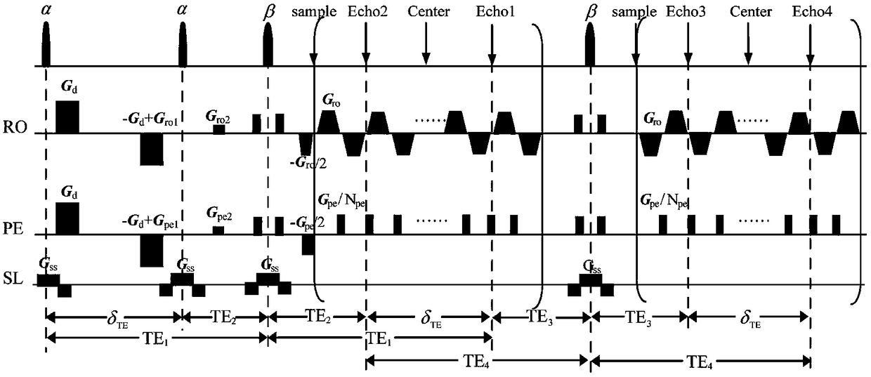
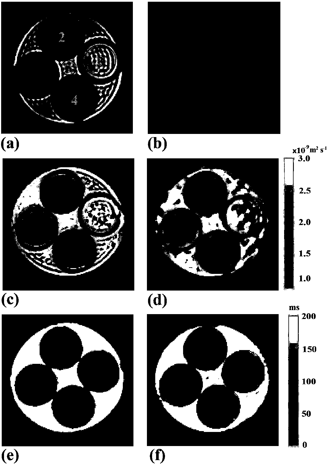
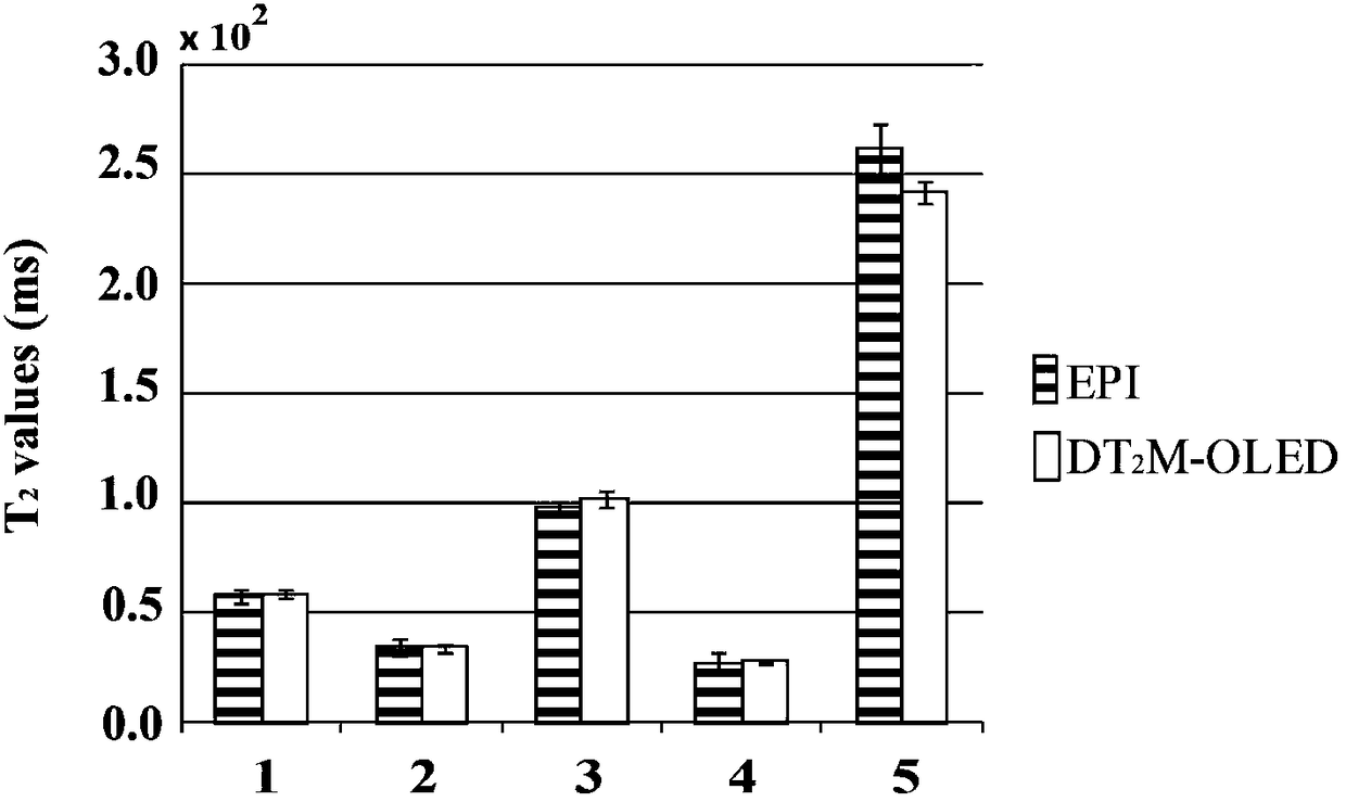
[0074] Using single-scan synchronous magnetic resonance diffusion and T based on overlapped echo 2 A model experiment was performed on the imaging method to verify the feasibility of the present invention. Before the experiment, water, tetrahydrate, manganese chloride (Mncl 2 .4H 2 O) and agarose are prepared according to the ratio in Table 1. figure 1 Model in the gel. The experiment was carried out under a small animal MRI 7T imager. On the operating table of the magnetic resonance imager, open the corresponding operating software in the imager, first locate the region of interest for the imaged object, and then perform tuning, shimming, power and frequency correction. In order to evaluate the effectiveness of the image obtained by this method, an Echo Planar Imaging (EPI) imaging experiment was carried out in the same environment as a comparison. Then import the compiled DT 2 M-OLED imaging sequence (e.g. figure 1 ), according to the specific experimental conditions, set ...


PUM


Abstract

A single-scan synchronous magnetic resonance diffusion and T2 imaging method based on overlapping echoes, relates to magnetic resonance imaging. Four echoes are produced by using a small angle excitation pulse and two echo chain samplings with two identical deflection angles, and each echo chain collects two echo signals. There is an evolution time and a pair of diffusion gradients after the firstexcitation pulse, due to which the transverse relaxation times and diffusion weights of the two echo signals in the first sampling are different. A shifting gradient of frequency dimension and phasedimension is added after each excitation pulse so that the signals generated by the different excitation pulses are different in position in the k-space. Two echo signals are re-converged with one refocusing pulse after finishing the first sampling, and then the second sampling is performed, in which two echo signals with the same transverse relaxation time and different diffusion weights can be obtained. The sampled signal is reconstructed using deep learning to obtain quantitative T2 and ADC images, and T2 and ADC images can be obtained in a single-scan.

Description

Technical field [0001] The present invention relates to magnetic resonance imaging, in particular to a single scan synchronous magnetic resonance diffusion (apparent diffusion coefficient, ADC) and T 2 Imaging method. Background technique [0002] Quantitative magnetic resonance imaging can provide a non-invasive way to analyze normal and diseased living tissues. It has been applied to the study of the microstructure of the brain, heart, and spinal cord. [1,2] . Quantitative imaging can eliminate the effects that are independent of the nature of the tissue, such as dependence on the operator, differences in scanning parameters, spatial changes in magnetic fields, and image scaling. [3] . Multi-parameter quantitative imaging is gaining more and more attention in clinical magnetic resonance imaging (MRI). Multi-parameter quantitative imaging acquires quantitative maps of different parameters (such as T 2 And diffusion maps), and use these maps to perform a joint analysis on the re...

Claims


Application Information

Patent Timeline
Patent Type & Authority Applications(China)
IPC IPC(8): G01R33/50G01R33/54G01R33/563A61B5/055
CPCA61B5/055G01R33/50G01R33/54G01R33/56341
Inventor 蔡聪波马崚嶒陈忠蔡淑惠丁兴号
Owner XIAMEN UNIV
Why Eureka
Try Eureka