Multi-color immunofluorescence labelling method and imaging method
What is AI technical title?
AI technical title is built by Patsnap AI team. It summarizes the technical point description of the patent document.
A technology of immunofluorescence and labeling method, applied in the field of immunofluorescence, which can solve the problems of color shifting and limited staining types, etc., and achieve the effect of clear colors
Pending Publication Date: 2019-02-01
姜云瀚
View PDF14 Cites 7 Cited by
Summary
Abstract
Description
Claims
Application Information
AI Technical Summary
This helps you quickly interpret patents by identifying the three key elements:
Problems solved by technology
Method used
Benefits of technology
Problems solved by technology
[0003] However, the staining types of the existing immunofluorescence techniques are limited, and when there are many staining types for a sample, color shifting is prone to occur
Method used
Image
Click on the blue label to locate the original text in one second.
Reading with bidirectional positioning of images and text.
Examples
Embodiment 1
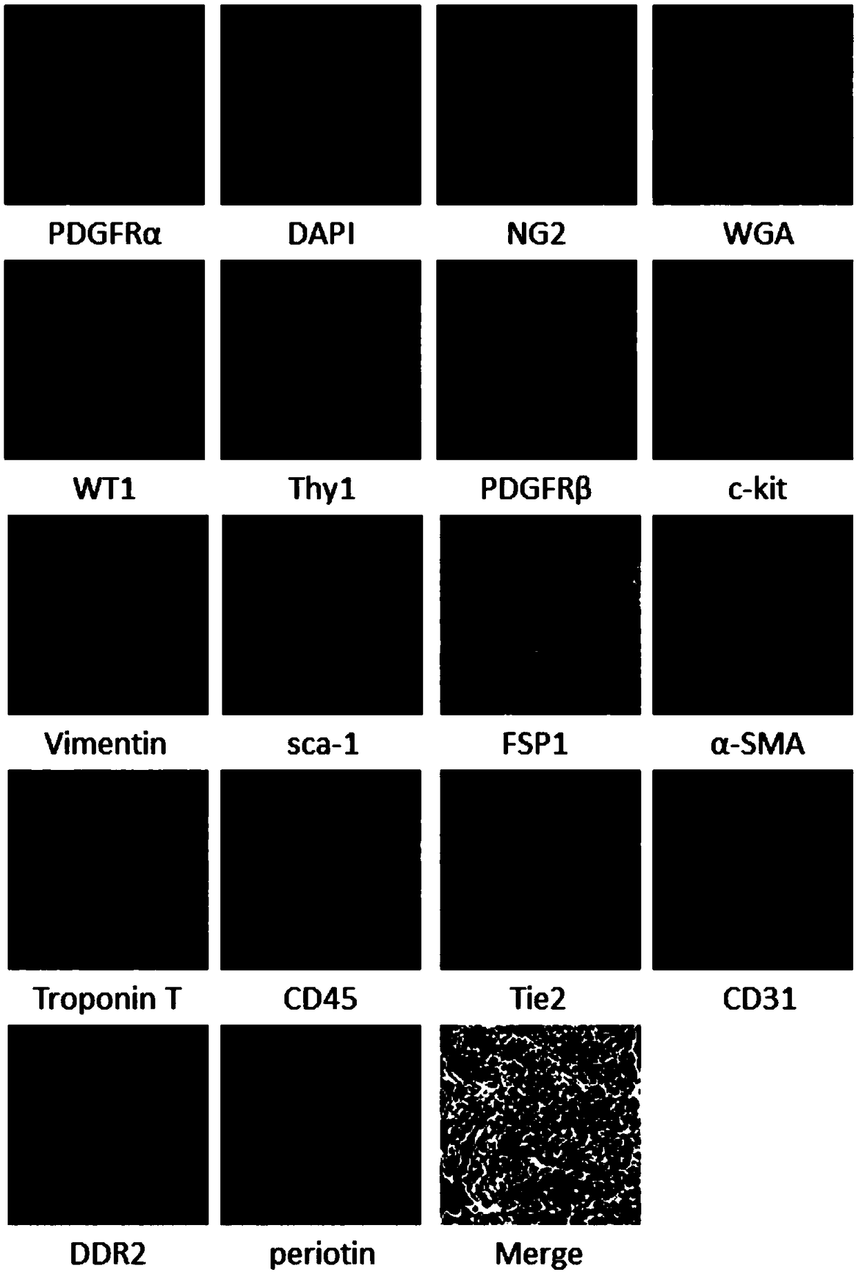
[0046] In this embodiment, the frozen section of C57 / B6 mouse heart is taken as a sample, and the multicolor immunofluorescent labeling method of this embodiment is described, as follows:
[0047] 1. Pretreatment
[0048] 1. Frozen sections of 1C57 / B6 mouse hearts were fixed with 4% paraformaldehyde for 15 minutes, and then washed with PBS buffer three times, 5 minutes each time.
[0049] 1.2 After washing, the frozen sections were incubated at 37°C for 1.5h with blocking membrane permeabilization solution.
[0050] Wherein, the sealing permeation solution is goat serum, which contains 0.3% Triton-100.
[0051] 2. The first fluorescent labeling
[0052] 2.1 Primary antibody incubation: use mouse-derived Troponin T antibody (purchased from Abcam, specification 200 μg, catalog number ab8295) (diluted at a ratio of 1:10) and rabbit-derived α-SMA protein-binding α -SMA antibody (purchased from Abcam, specification 10ul, product number ab32575) (diluted at a ratio of 1:20) and fro...
Embodiment 2
[0124] This embodiment provides a multi-color immunofluorescence imaging method, which uses the sample prepared in Embodiment 1 as the observation object, and uses a confocalmicroscope (Leica SP5 confocalmicroscopy) for imaging. The specific operation method is as follows:
[0126] Use 405 laser to illuminate the light of Dylight405 (take pictures in the range of 415-445nm);
[0127] Use a 405 laser to perform spectral scanning (scanning the wavelength range of 415-481nm, with 3nm as a scanning unit, and scan once every 1nm increase (the way of spectral scanning is the same)) (split Dylight405 and DAPI) (abandon Dylight405, Save DAPI's graph);
[0128] Use 405 laser to illuminate the light of Qdot625 (take pictures in the range of 600-660nm);
[0129] Use 488 laser to perform spectral scanning (scanning 500-548nm) (split AF488, PE-Cy7 and BODIPY FL);
[0130] Use 514 laser to perform spectral scanning (scanning 524-599nm) (split AF514, AF532, ...
PUM
Abstract
The invention discloses a multi-color immunofluorescencelabelling method and an imaging method, and relates to the technical field of immunofluorescence. The multi-color immunofluorescence labellingmethod comprises a plurality of fluorescencelabelling steps and crosslinking fixing steps of incubating a sample with a crosslinking fixative after each fluorescent labelling step. The crosslinking fixative comprises acetone, paraformaldehyde and PBS buffer. According to the multi-color immunofluorescence labelling method, various kinds of different fluorescent dyes can be labelled on various kinds of antigen markers in the sample. The labelled sample can be used for confocalmicroscopy imaging to display various kinds of different colors, and the colors are clear, bright and uncolored. The method provides a basis for scientific research workers to simultaneously observe the properties, positioning and content of various kinds of antigens.
Description
technical field [0001] The invention relates to the technical field of immunofluorescence, in particular to a multicolor immunofluorescence labeling method and an imaging method. Background technique [0002] Immunofluorescent staining technology is based on the principle of antigen-antibody reaction. Firstly, known antigens or antibodies are labeled with fluorescein to make fluorescent markers, and then this fluorescent antibody (or antigen) is used as a molecular probe to examine cells or tissues. Corresponding antigen (or antibody). The antigen-antibody complexes formed in cells or tissues contain fluorescein. Use a fluorescencemicroscope to observe the specimen. The fluorescein emits bright fluorescence (yellow-green or orange-red) under the irradiation of excitation light, and the cells or tissues where the fluorescence is located can be seen. , so as to determine the nature and location of the antigen or antibody, and use quantitative techniques to determine the cont...
Claims
Application Information
Patent Timeline
Application Date:The date an application was filed.
Publication Date:The date a patent or application was officially published.
First Publication Date:The earliest publication date of a patent with the same application number.
Issue Date:Publication date of the patent grant document.
PCT Entry Date:The Entry date of PCT National Phase.
Estimated Expiry Date:The statutory expiry date of a patent right according to the Patent Law, and it is the longest term of protection that the patent right can achieve without the termination of the patent right due to other reasons(Term extension factor has been taken into account ).
Invalid Date:Actual expiry date is based on effective date or publication date of legal transaction data of invalid patent.

