
Detecting method based on coaxial time-domain distinguishing photoacoustic imaging

A detection method and photoacoustic imaging technology, applied in diagnostic recording/measurement, medical science, sensors, etc., to reduce measurement errors

Active Publication Date: 2019-02-22
SHANGHAI INST OF TECHNICAL PHYSICS - CHINESE ACAD OF SCI
View PDF4 Cites 2 Cited by
  • Summary
  • Abstract
  • Description
  • Claims
  • Application Information

AI Technical Summary

Problems solved by technology

Photoacoustic imaging technology can realize the detection of blood vessel distribution, that is, the detection of external shape, but it is a difficult problem to be solved to detect the external shape of blood vessels and the distribution of internal tissue morphology at the same time.

Method used


Image

  • Detecting method based on coaxial time-domain distinguishing photoacoustic imaging

Examples


Embodiment Construction

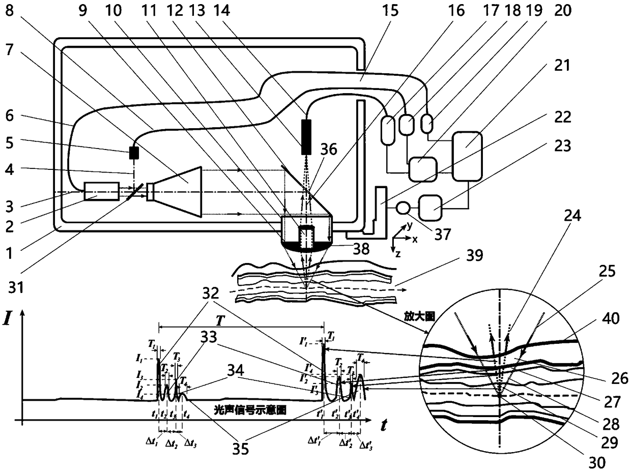
[0029] The specific embodiment of the present invention is as figure 1 shown.

[0030] The human blood vessel detection method based on laser photoacoustic proposed by the present invention is implemented on a laser photoacoustic based human blood vessel detection system, which mainly consists of a main controller 21, a scanning head 1, a three-dimensional electric platform 22 and an auxiliary component composition;

[0031] Wherein, auxiliary components include cascade amplifier 17, detector circuit 18, laser controller 19, signal acquisition card 20, three-way motor controller 23 and three-way stepper motor 37;

[0032] Inside the scanning head 1 are a photoacoustic probe 10, a perforated mirror 12, a laser 2, a laser beam expander 7, a proportional beam splitter 31, a photoelectric detector 5, an ultrasonic probe 13, a laser cable 6, and a detector cable 8 and an ultrasonic cable 14; a window 15 is provided on the scanning head 1, so that the laser cable 6, the detector c...


PUM


Abstract

The invention discloses a detecting method based on coaxial time-domain distinguishing photoacoustic imaging. The method includes the steps of system initial positioning, single-point testing, time-domain distinguishing photoacoustic positioning and analyzing, photoacoustic single-point blood vessel measuring and photoacoustic three-dimensional blood vessel reestablishing. The detecting method hasthe advantages that through a coaxial laser emitting and ultrasonic receiving light path, an optical focus and an acoustic focus coincide, same-vortex laser stereoscopic angle exciting and receivingstereoscopic angle receiving are achieved, and measurement errors can be reduced; with blood flow photoacoustic echoes in blood vessels as the space analysis standard, through time-domain distinguishing photoacoustic echo signal analysis, photoacoustic echo signals corresponding to the blood flow, blood vessel inner tissue and blood vessel wall layered substances passing through a main-axis routeat the same time can be analyzed, and photoacoustic detection can be conducted on the outer appearance of the blood vessels and the inner tissue form distribution at the same time.

Description

technical field [0001] The invention relates to a human blood vessel detection method, in particular to a detection method based on coaxial time-domain resolution photoacoustic imaging, which is suitable for detection of three-dimensional structures inside and outside human arteries and veins, and belongs to the field of photoelectric detection. Background technique [0002] Angiography is an auxiliary inspection technique used to monitor the health of blood vessels. It is widely used in the diagnosis and treatment of various clinical diseases. It helps doctors to detect the disease in time, control the progress of the disease, and effectively improve the survival of patients. Rate. Angiography is an interventional detection method. It needs to inject a contrast agent into the blood vessels of the subject. Because X-rays cannot penetrate the contrast agent, angiography can accurately reflect the location and degree of vascular lesions. However, angiography also has many def...

Claims


Application Information

Patent Timeline
IPC IPC(8): A61B5/00A61B5/02
CPCA61B5/0095A61B5/02007
Inventor 万雄王泓鹏袁汝俊何强
Owner SHANGHAI INST OF TECHNICAL PHYSICS - CHINESE ACAD OF SCI
Learn More