
Real time dosimetry of ultrasound imaging probe

A technology of ultrasonic probe and dosimeter, which is applied in ultrasonic/acoustic/infrasonic diagnosis, acoustic diagnosis, infrasonic diagnosis, etc., to achieve the effect of improving quality control

Active Publication Date: 2019-03-01
KONINKLJIJKE PHILIPS NV
View PDF11 Cites 0 Cited by
  • Summary
  • Abstract
  • Description
  • Claims
  • Application Information

AI Technical Summary

Problems solved by technology

However, the use of US imaging during the delivery portion of the procedure presents certain difficulties

Method used


Image

  • Real time dosimetry of ultrasound imaging probe
  • Real time dosimetry of ultrasound imaging probe
  • Real time dosimetry of ultrasound imaging probe

Examples


Embodiment Construction

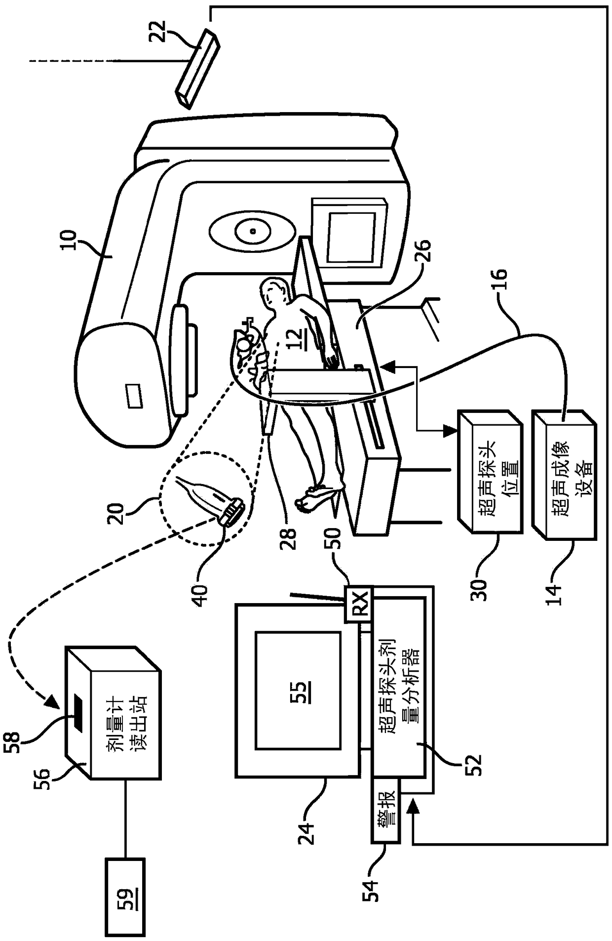
[0022] Radiation therapy sessions employing an external radiation beam and including ultrasound (US) monitoring should be designed to avoid irradiating the US probe with the therapeutic ionizing radiation used to deliver the radiation therapy. This can be achieved by placing the US probe outside the beam path but close enough to the irradiated area of ​​the subject to provide useful US imaging.

[0023] However, it is recognized herein that some radiation exposure from US probes cannot always be avoided, especially in the context of multibeam or tomographic radiation therapy where a single fixed radiation beam path does not exist. Even if the period is designed to avoid irradiating the US probe, errors may occur in placement of the US probe, placement of the patient, or other types of errors that may result in radiation exposure of the US probe. Furthermore, even if the US probe never enters the path of the radiation beam during a radiation treatment session, stray ionizing ra...


PUM


Abstract

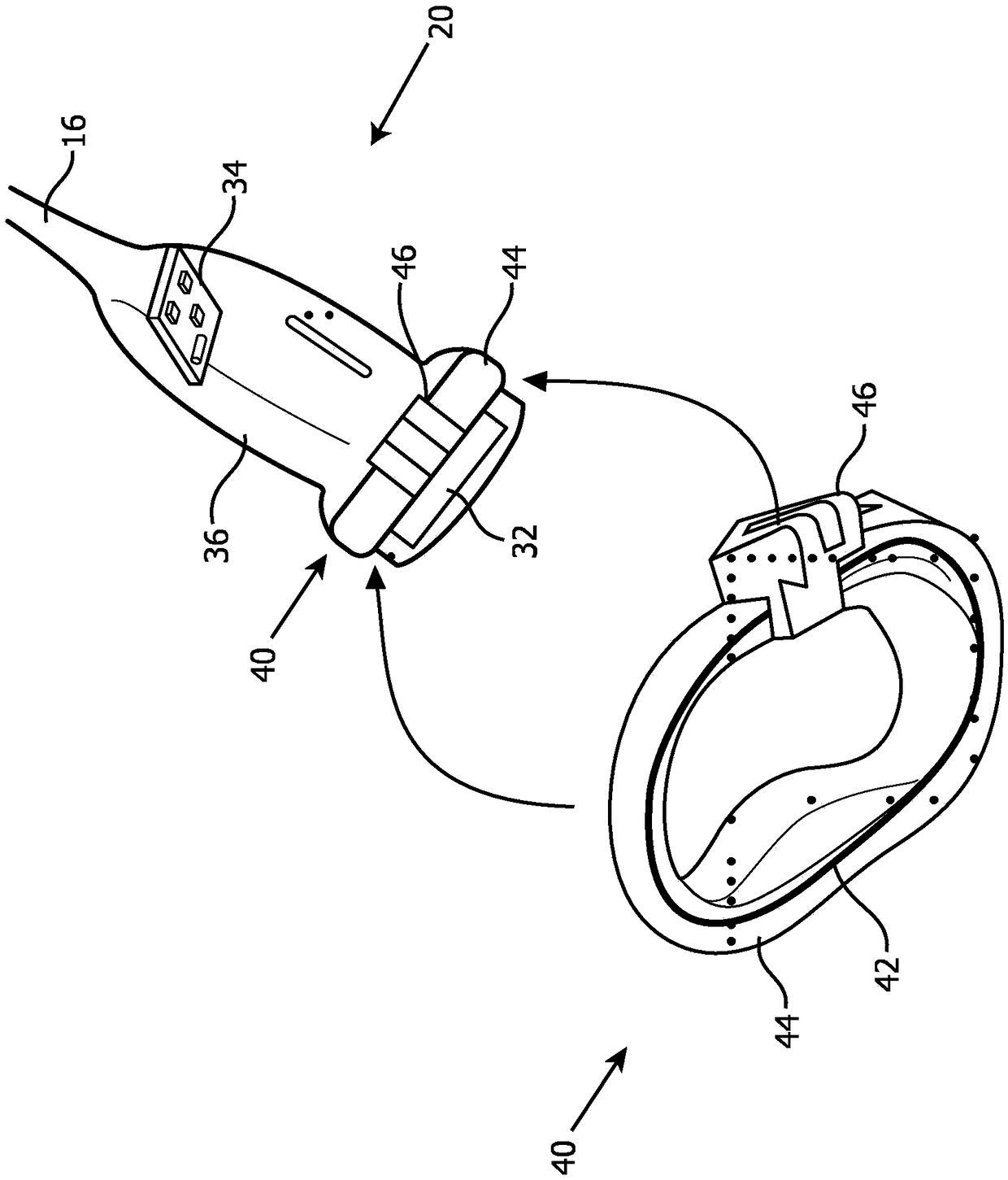
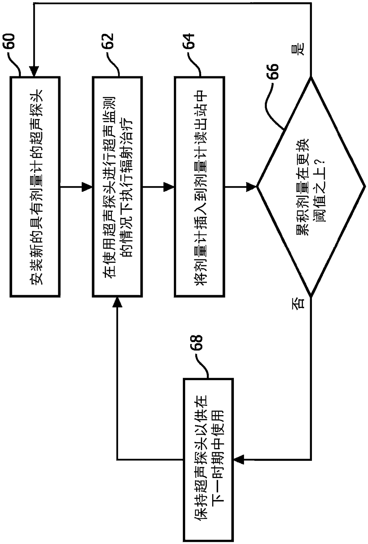
An ultrasound probe includes an ultrasound probe housing (36) and one or more ultrasound transducers (32) disposed in the ultrasound probe housing. A dosimeter (40) or ionizing radiation detector is disposed in or attached to the ultrasound probe housing. An alarm device (24, 52, 54, 56) receives radiation dose or radiation exposure data acquired by the dosimeter or ionizing radiation detector. The alarm device includes an electronic processor (24, 56) programmed to detect excessive radiation dose or radiation exposure received by the ultrasound probe based on the radiation dose or radiation exposure data acquired by the dosimeter or ionizing radiation detector, and output an alarm warning of the detection of excessive radiation dose or radiation exposure received by the ultrasound probe.In some embodiments, the dosimeter is a one-time use dosimeter that is not resettable.

Description

technical field [0001] The following relates generally to the field of external beam radiation therapy (EBRT), the field of ultrasound procedure monitoring, and the like. Background technique [0002] External beam radiation therapy (EBRT) uses beams of ionizing radiation as an oncological treatment tool. The ionizing radiation may be an electron beam, a proton beam, an X-ray beam, or the like. EBRT is typically delivered in a fractionated treatment approach that involves multiple phases beginning with pre-treatment imaging, followed by image-based treatment planning, and ending with multiple treatment delivery. Deliver each portion every other day. [0003] A difficulty with fractionated EBRT is that the target tumor and / or radiosensitive critical organs may shift between treatment delivery segments (inter-segment motion) or even during a single treatment delivery segment (intra-segment motion). Monitoring such motion can be problematic, especially in the case of motion ...

Claims


Application Information

Patent Timeline
IPC IPC(8): A61N5/10
CPCA61N2005/1058A61N2005/1094A61N5/1049A61B8/4218A61B8/4444A61B8/4472A61B8/4483A61N5/1071A61N2005/1087A61N2005/1089A61N2005/1095
Inventor V·帕塔萨拉蒂A·L·鲁滨逊
Owner KONINKLJIJKE PHILIPS NV
Learn More