An intelligent medical image segmentation method based on three-dimensional reconstruction

A medical imaging and three-dimensional reconstruction technology, applied in image analysis, image enhancement, image data processing, etc., can solve the problems of not considering image position information, difficult to meet segmentation accuracy, difficult to obtain segmentation results, etc., to reduce the amount of calculation and operation. Time, good segmentation effect, and the effect of ensuring accuracy

Active Publication Date: 2019-03-22
SOUTH CHINA UNIV OF TECH
View PDF3 Cites 47 Cited by
  • Summary
  • Abstract
  • Description
  • Claims
  • Application Information

AI Technical Summary

Problems solved by technology

[0008] 1. Using prior knowledge for segmentation, for complex medical images, any single segmentation algorithm is difficult to obtain satisfactory segmentation results
[0009] 2. Most of the segmentation algorithms are based on two-dimensional image data, without considering the position

Method used


Image

  • An intelligent medical image segmentation method based on three-dimensional reconstruction
  • An intelligent medical image segmentation method based on three-dimensional reconstruction
  • An intelligent medical image segmentation method based on three-dimensional reconstruction

Examples


Embodiment Construction

[0059] The present invention will be further described below in conjunction with specific examples.

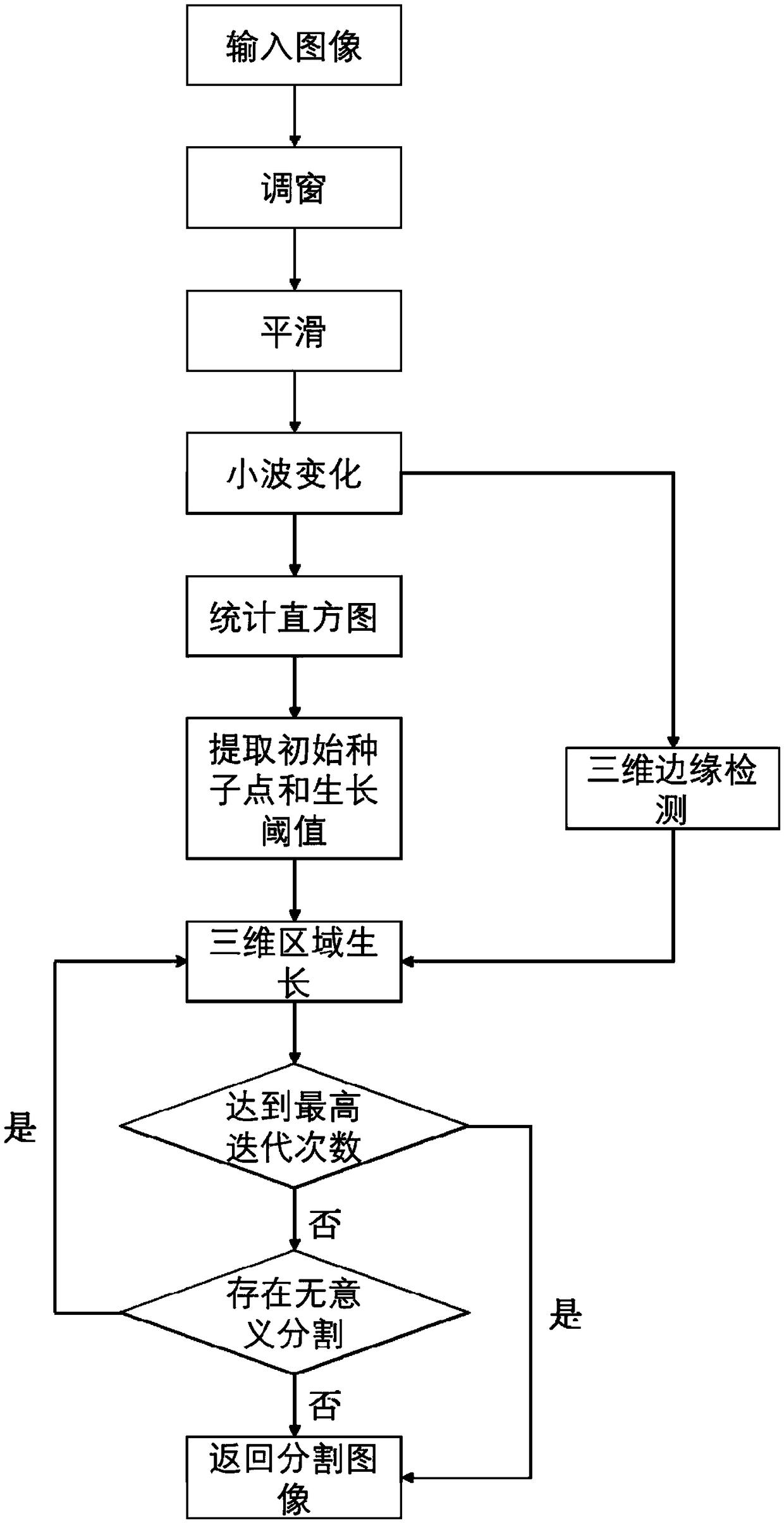
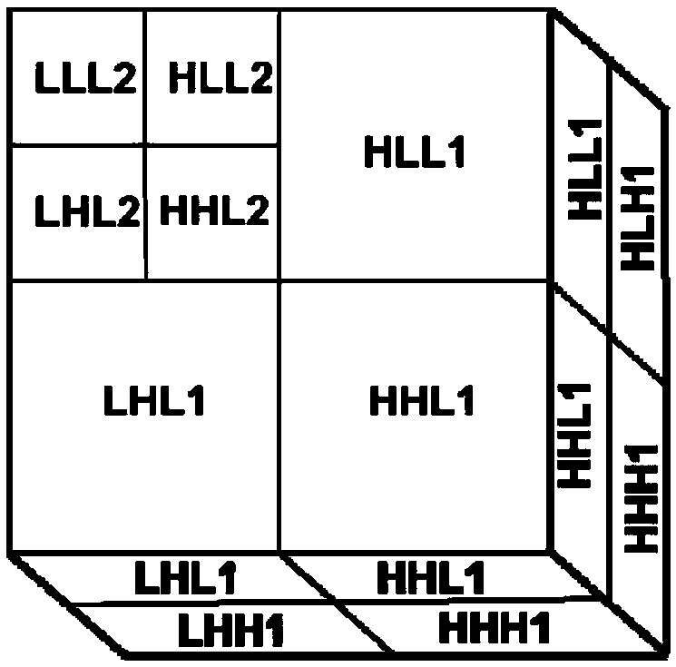
[0060] Such as figure 1 As shown, the intelligent segmentation method of medical image tissue based on three-dimensional reconstruction provided by the present invention includes the following steps:

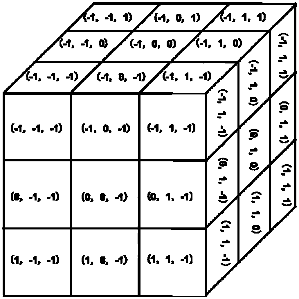
[0061] The first step is to input a set of medical image sequences in DICOM format, and perform windowing and Gaussian smoothing on the image sequences in the preprocessing stage. The so-called window adjustment is to convert the gray value in the window in the image data to the value in the brightest and darkest range when displayed according to the predicted window width and window level, and set the part higher than the gray range of the window to the lowest. Bright, and the part below the grayscale range of the window is set to the darkest. The window adjustment formula is as follows:

[0062]

[0063] Among them, F' xyz is the gray value of the pixel at the three-dim...


PUM


Abstract

The invention discloses an intelligent segmentation method of medical image organization based on three-dimensional reconstruction, which comprises the following steps: 1) inputting DICOM medical image sequence, preprocessing the image and adjusting the window; 2) performing wavelet transformation on that image to reduce the image data to 1/4 of the original image; 3) performing statistic gray histogram to extract initial seed point and growth threshold; 4) carrying out three-dimensional edge detection on that image to obtain an edge contour map; 5) carrying out three-dimensional space regiongrowth by combining gray level and edge information; 6) Calculating the region attributes after segmentation, optimize the seed point and growth threshold. Through statistical histogram, the inventioncan automatically extract initial seed points required for region growth and threshold required for growth criterion, and then carry out three-dimensional region growth combined with edge contour information of an image, and continuously optimize initial seed points and growth threshold through iterative growth, thereby improving segmentation results. At that same time, the invention reduces theimage data to 1/4 of the original image through the wavelet change, thereby shortening the operation time.

Description

technical field [0001] The invention relates to the technical field of medical image segmentation, in particular to an intelligent segmentation method of medical image tissue based on three-dimensional reconstruction. Background technique [0002] Medical image segmentation refers to separating regions with different meanings in medical image data according to a specific property, so that the same regions meet regional consistency. Commonly used medical image segmentation methods include: threshold-based segmentation methods, region-based segmentation methods, edge-based segmentation methods, and cluster-based segmentation methods. [0003] The threshold-based segmentation method is to use one or a group of gray thresholds to mark the image data within the threshold range as the region of interest, and mark the image data outside the range as background data, so as to achieve the purpose of image segmentation. The threshold-based segmentation method has the advantages of si...

Claims


Application Information

Patent Timeline
IPC IPC(8): G06T7/13G06T5/00G06T5/40
CPCG06T5/002G06T5/40G06T2207/10021G06T2207/30004G06T7/13
Inventor 许洁斌丁晓芳陈家兴
Owner SOUTH CHINA UNIV OF TECH
Why Eureka
Try Eureka