Semantics-based medical imaging report template generation method

A medical image and report template technology, applied in the field of medical image processing, can solve the problem of cumbersome and time-consuming image report writing.

Pending Publication Date: 2019-03-29
FUDAN UNIV +1
View PDF0 Cites 20 Cited by
  • Summary
  • Abstract
  • Description
  • Claims
  • Application Information

AI Technical Summary

Problems solved by technology

For inexperienced radiologists, the requirements for writing image reports are very high. For example, to be able to read chest X-ray images correctly, it is necessary to understand the anatomy of the chest cavity, the physiological characteristics of chest disease

Method used


Image

  • Semantics-based medical imaging report template generation method
  • Semantics-based medical imaging report template generation method
  • Semantics-based medical imaging report template generation method

Examples


Example Embodiment

[0049] The following is a report of chest X-ray images to show the specific implementation of this method:

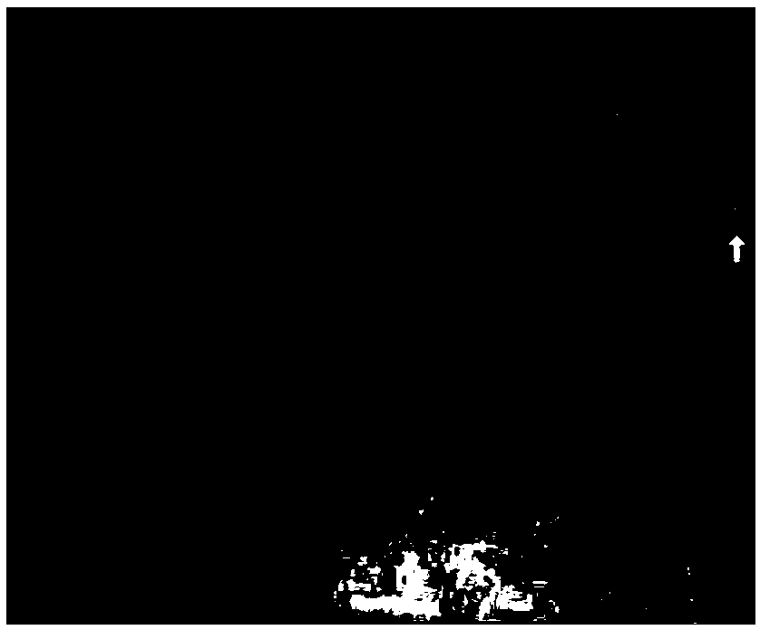
[0050] (1) Input image see figure 2 As shown, the pathological labels actually contained in the image are "bilateral pleuraleffusion", "degenerative joint disease", and "pleural effusion", and the actual content of the report is

[0051] "Small bilateral pleural effusions.Prominent interstitialmarkings.There are small bilateral pleural effusions.No pneumothorax or focalconsolidation.Normal heart size.Catheter tubing present in the uppermidabdomen.There is bilateral acromioclavicular degenerative joint disease, right greater than left."

[0052] (2) The image is input into the trained VGG19 network, and 512*14*14 features are extracted

[0053] (3) After the feature global pooling is input to the multi-label prediction module, the pathological label probability is output, and the labels of the probability value top-5 are "congestive heart failure", "edemas", "degenerative joint...


PUM


Abstract

The invention belongs to the technical field of medical image processing and particularly relates to a semantics-based medical imaging report template generation method. An initial imaging report is generated automatically on the basis of the image, so that a reference template is provided for inexperienced roentgenologists to write imaging reports and the working strength and the difficulty of the roentgenologists are reduced. When writing the image reports, the roentgenologists can pay much attention to different areas according to a certain diagnosis sequence and can make corresponding imaging description according to the characteristics of the areas. An image encoder and hierarchical sentence decoder framework is used, attention mechanism according to reference text is introduced, andthe process of writing the imaging reports by the professional roentgenologists is simulated. Compared with the traditional template-based and rule-based method, the method provided by the invention has the advantages of generating natural language imaging reports and capturing rich semantic characteristics according to the image characteristics.

Description

technical field [0001] The invention belongs to the technical field of medical image processing, and in particular relates to a method for generating a medical image report template based on semantics. Background technique [0002] With the development of image imaging technology, medical images such as X-ray (X-ray) images, (Computed Tomography, computerized tomography) images, MR (Magnetic Resonance, nuclear magnetic resonance) images, etc. have been widely used in hospitals and clinics, and can be used in Including the screening and diagnosis of pneumonia, pneumothorax, pulmonary nodules, esophageal cancer, breast cancer and other diseases, providing more and more support for subsequent clinical diagnosis. Radiologists read the images, describe findings in various regions of the images, and write text reports. For inexperienced radiologists, the requirements for writing image reports are very high. For example, to be able to read chest X-ray images correctly, it is neces...

Claims


Application Information

Patent Timeline
IPC IPC(8): G16H15/00G16H30/00
CPCG16H15/00G16H30/00
Inventor 熊贇谢贤成李康安朱扬勇
Owner FUDAN UNIV
Why Eureka
Try Eureka