mammary gland MRI lesion area detection method based on multi-dimensional information fusion

A lesion area and detection method technology, which is applied in image data processing, image enhancement, instruments, etc., can solve the problems of difficulty in distinguishing, inaccurate breast magnetic resonance image detection results, and low recall rate of target detection in lesion area, etc. The effect of classification accuracy and accurate detection

Pending Publication Date: 2019-04-19
泰格麦迪(北京)医疗科技有限公司
View PDF4 Cites 4 Cited by
  • Summary
  • Abstract
  • Description
  • Claims
  • Application Information

AI Technical Summary

Problems solved by technology

However, the image features of some diseased areas in the breast have a high degree of similarity with normal tissues (for example, blood vessel sections) in a single image, and it is difficult to effectively distinguish them using the above-mentioned prior art
Moreover, when the existing saliency d

Method used


Image

  • mammary gland MRI lesion area detection method based on multi-dimensional information fusion
  • mammary gland MRI lesion area detection method based on multi-dimensional information fusion
  • mammary gland MRI lesion area detection method based on multi-dimensional information fusion

Examples


Embodiment Construction

[0019] In order to make the object, technical solution and advantages of the present invention clearer, the present invention will be further described in detail below in conjunction with the accompanying drawings.

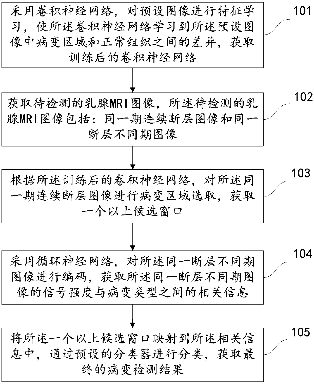
[0020] In the present invention, we adopt the algorithm based on candidate regions in natural image target detection, and propose to use convolutional neural network to extract breast MRI image information of different sections and different time sequences, and use images of different sections mainly for the extraction of candidate regions. A method for further classifying and judging candidate regions by using images of different time series.

[0021] figure 1 It is a flow chart of a method in an embodiment of the present invention, specifically including the following steps:

[0022] Step 101, using the convolutional neural network to perform feature learning on the preset image, so that the convolutional neural network can learn the difference between the lesi...


PUM


Abstract

The invention discloses a mammary gland MRI lesion area detection method based on multi-dimensional information fusion, and the method comprises the steps: carrying out the feature learning of a preset image through employing a convolutional neural network, and enabling the convolutional neural network to learn the difference between a lesion area and a normal tissue in the preset image; Acquiringa mammary gland MRI image to be detected, wherein the mammary gland MRI image comprises a same-period continuous tomography image and a same-fault different-period image; According to the trained convolutional neural network, lesion area selection is carried out on the continuous tomography images of the same period, and more than one candidate window is obtained; Encoding the images of differentperiods of the same fault by adopting a recurrent neural network to obtain relevant information between the signal intensity and the lesion type of the images of different periods of the same fault;And mapping more than one candidate window into the related information, and classifying through a preset classifier to obtain a final lesion area detection result. According to the technical scheme provided by the invention, the lesion area in the mammary gland can be more accurately detected.

Description

technical field [0001] The invention relates to the technical field of magnetic resonance imaging, in particular to a method for detecting breast MRI lesion regions based on multi-dimensional information fusion. Background technique [0002] Magnetic resonance imaging (Magnetic Resonance Imaging, MRI) technology is a technology that uses the nuclear magnetic resonance phenomenon of hydrogen protons for imaging. With the continuous maturity of magnetic resonance imaging technology in recent years, its importance in the diagnosis of breast diseases is increasing. Breast magnetic resonance technology is more sensitive to the further judgment of the nature of breast lesions. It can not only make further judgments on the benign and malignant lesions, but also help doctors identify whether there are multiple lesions. In particular, breast magnetic resonance technology can make a more accurate judgment on the benign and malignant lesions according to the change of the signal inten...

Claims


Application Information

Patent Timeline
IPC IPC(8): G06T7/00G06N3/04
CPCG06T7/0012G06T2207/30068G06T2207/10088G06N3/044G06N3/045
Inventor 陈小刚
Owner 泰格麦迪(北京)医疗科技有限公司
Why Eureka
Try Eureka