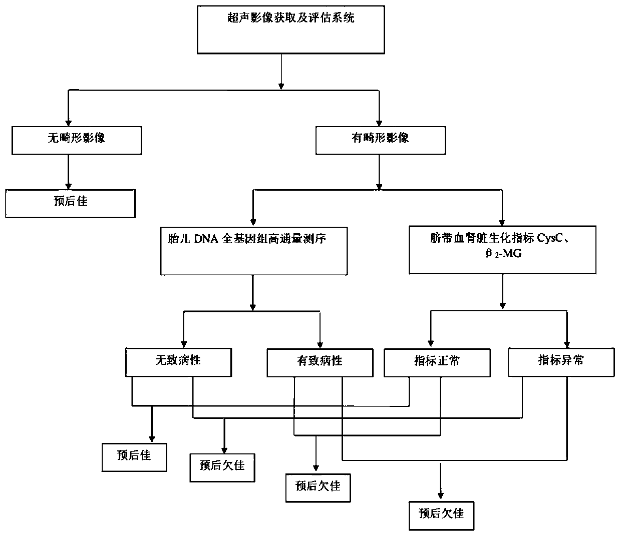
Method for fetal urinary system malformation comprehensive evaluation
A comprehensive evaluation and technology of the urinary system, applied in urinary function evaluation, ultrasound/sonic/infrasonic Permian technology, ultrasound/sonic/infrasonic image/data processing, etc., can solve the problem of not being able to provide information about the cause of disease, kidney function, etc. question
- Summary
- Abstract
- Description
- Claims
- Application Information
AI Technical Summary
Problems solved by technology
Method used
Image
Examples
experiment example 1
[0030] Pregnant woman A inspection time: 23 weeks pregnant on May 24, 2018
[0031] 1. Ultrasound image acquisition and evaluation system: Use a color Doppler ultrasound instrument with a probe frequency of 3.5MHz to obtain the position of the kidneys, the size of the kidneys, the renal artery, the circumference of the kidneys / abdominal circumference, and the kidneys of the fetus in the pregnant woman's abdomen. Morphological characteristics such as cortical echo, cortical thickness, bilateral renal pelvis separation, bilateral ureter expansion, bladder size, shape, and amniotic fluid index. According to different contents, different image pictures are collected and stored in the ultrasound workstation. Ultrasound images show that the amniotic fluid index is 1.0 cm, and it is assessed as oligohydramnios, and the fetal urinary system may be deformed, so follow-up evaluation is performed.
[0032]2. After the step (1) outputs fetal urinary system abnormalities, interventional s...
experiment example 2
[0047] Pregnant woman B examination time: 23 weeks pregnant on July 19, 2018
[0048] 1. The method is the same as that of Experiment 1. Ultrasound image acquisition and evaluation system: Use a color Doppler ultrasound instrument with a probe frequency of 3.5MHz to obtain the position of the kidneys, the size of the kidneys, the renal artery, the circumference / abdominal circumference of the kidneys, and the cortex and medulla of the kidneys of the fetus in the pregnant woman's abdomen Morphological characteristics such as echo, cortical thickness, separation of bilateral renal pelvis, dilatation of bilateral ureter, bladder size, shape, and amniotic fluid index. According to different contents, different image pictures are collected and stored in the ultrasound workstation. Ultrasound images showed that the fetal left kidney was 1.4x1.0cm in size, the right kidney was 1.2x0.9cm in size, and the amniotic fluid index was 4.3cm. Ultrasound images show that if the kidneys are u...
PUM
 Abstract
Description
Claims
Application Information
 - R&D
- Intellectual Property
- Life Sciences
- Materials
- Tech Scout
- Unparalleled Data Quality
- Higher Quality Content
- 60% Fewer Hallucinations
Browse by: Latest US Patents, China's latest patents, Technical Efficacy Thesaurus, Application Domain, Technology Topic, Popular Technical Reports.
© 2025 PatSnap. All rights reserved.Legal|Privacy policy|Modern Slavery Act Transparency Statement|Sitemap|About US| Contact US: help@patsnap.com

