Confocal microscopic imaging system and method in vacuum cavity based on cage structure

A technology of confocal microscopy and cage structure, applied in microscopes, optics, instruments, etc., can solve the problems of too many optical elements, large space occupation, complex structure, etc., and achieve high control accuracy, high stability, and improved The effect of the signal-to-noise ratio

Pending Publication Date: 2019-05-31
UNIV OF SCI & TECH OF CHINA
View PDF4 Cites 3 Cited by
  • Summary
  • Abstract
  • Description
  • Claims
  • Application Information

AI Technical Summary

Problems solved by technology

[0003] At present, the microscopic imaging systems equipped with commercial vacuum chambers or cryogenic chambers generally have a relatively complex structure, occupy a large space, and have too many optical components, thereby reducing the collection efficiency and detection efficiency of fluorescent signals, and the cost is generally high.

Method used


Image

  • Confocal microscopic imaging system and method in vacuum cavity based on cage structure
  • Confocal microscopic imaging system and method in vacuum cavity based on cage structure
  • Confocal microscopic imaging system and method in vacuum cavity based on cage structure

Examples


Embodiment Construction

[0051] The technical solutions in the embodiments of the present invention will be clearly and completely described below in conjunction with the accompanying drawings in the embodiments of the present invention. Obviously, the described embodiments are only some of the embodiments of the present invention, not all of them. Based on the embodiments of the present invention, all other embodiments obtained by persons of ordinary skill in the art without making creative efforts belong to the protection scope of the present invention.

[0052] According to attached Figure 1-6 Shown, the present invention is further described.

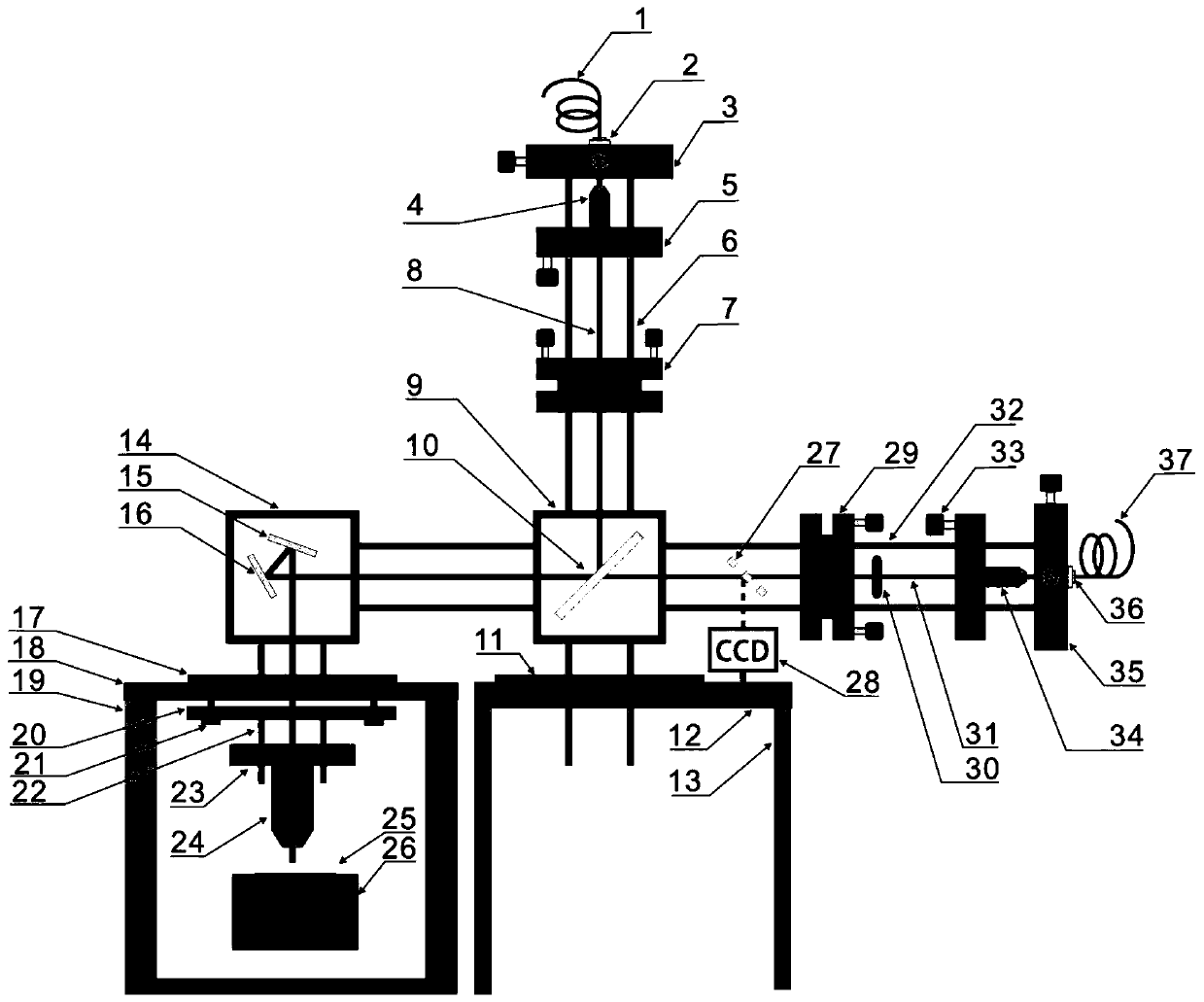
[0053] figure 1 It is a schematic diagram of the device structure provided by an embodiment of the present invention.

[0054] The upper right side of the microscope objective lens is a fluorescent microscope barrel with a cage structure, which is composed of a laser incident module in a vertical direction and a fluorescence collecting module in a horizo...


PUM


Abstract

The invention discloses a confocal microscopic imaging system and method in a vacuum cavity based on a cage structure. The system comprises a vacuum cavity, a vacuum cavity sealing cover, a quartz window piece, a three-dimensional electric displacement stage, a galvanometer system, a microscope objective, a fluorescence microscope tube, a cage structure optomechanical module, a laser, a single mode fiber, a fiber splitter, a dichroic mirror, an optical filter, an imaging CCD, a single photon detector, a pulse counter, a calibrator and a computer. The three-dimensional electric displacement stage and the microscope objective are arranged in the vacuum cavity, the galvanometer system and the main body of the microscope system are arranged at the outer portion of the vacuum cavity, and the main body of the microscope system employs a stable cage structure which is formed by the easily installed cage structure optomechanical module. The confocal microscopic imaging system and method are high in integration degree, small in occupied volume, easy to install and debugg and simple to operate and maintain, can be used for high-precision imaging of eak fluorescent signals in the vacuum cavity or the low-temperature cavity and high efficiency collection and signal analysis of a single photon source.

Description

technical field [0001] The invention relates to the field of optical microscopic imaging, in particular to a confocal microscopic imaging system and method in a vacuum cavity based on a cage structure. Background technique [0002] Confocal microscope is a common scientific research equipment for studying fluorescence imaging and weak fluorescence signal collection, detection and signal analysis. In some specific cases, researchers need to study the properties of fluorescence radiated by samples in vacuum or cryogenic environments. However, imaging, collecting, detecting and analyzing sample fluorescence in vacuum or cryogenic chambers usually requires The user builds the microscope imaging system by himself. [0003] At present, the microscopic imaging systems equipped with commercial vacuum chambers or cryogenic chambers generally have complex structures, occupy a large space, and have too many optical components, thereby reducing the collection efficiency and detection e...

Claims


Application Information

Patent Timeline
Patent Type & Authority Applications(China)
IPC IPC(8): G02B21/00G01N21/64
Inventor 李强王俊峰闫飞飞程则迪许金时李传锋
Owner UNIV OF SCI & TECH OF CHINA
Why Eureka
Try Eureka