A retinal fundus image cup/disc ratio automatic evaluation method

A fundus image, automatic evaluation technology, applied in image enhancement, image analysis, image data processing and other directions, can solve the problem of manual extraction, inability to accurately segment the optic disc and optic cup at the same time, and achieve the effect of enhancing accuracy and robustness

Inactive Publication Date: 2019-05-31
CENT SOUTH UNIV
View PDF6 Cites 31 Cited by
  • Summary
  • Abstract
  • Description
  • Claims
  • Application Information

AI Technical Summary

Problems solved by technology

It solves the problem that the existing optic disc and cup segmentation methods need to ...

Method used


Image

  • A retinal fundus image cup/disc ratio automatic evaluation method
  • A retinal fundus image cup/disc ratio automatic evaluation method
  • A retinal fundus image cup/disc ratio automatic evaluation method

Examples


Embodiment 1

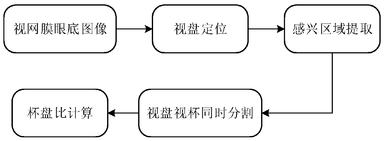
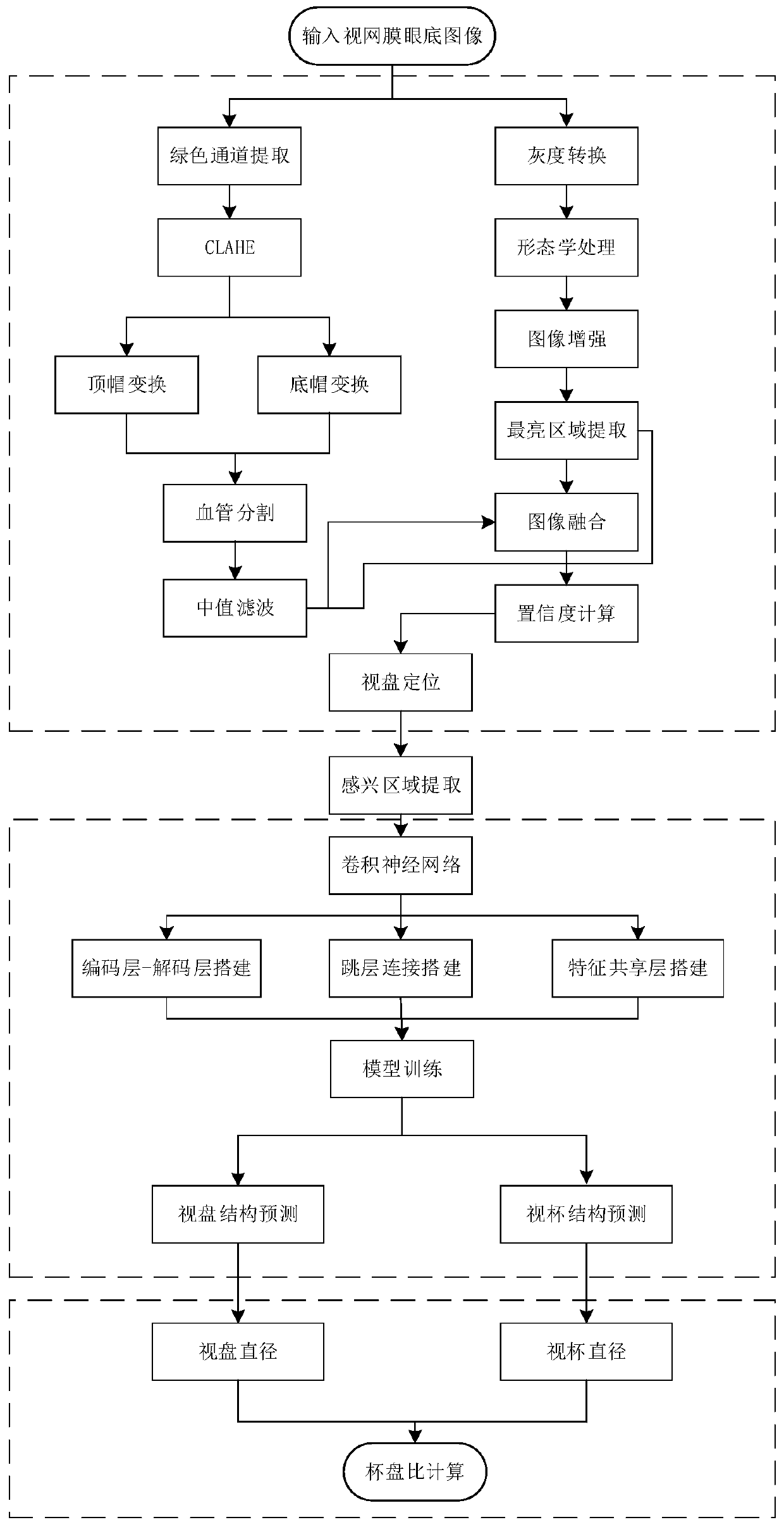
[0083] This embodiment is aimed at retinal fundus images, and the automatic evaluation of the cup-to-disk ratio is carried out in the following steps. The overall implementation process is as follows: figure 1 As shown, the detailed implementation process is as follows figure 2 Shown:

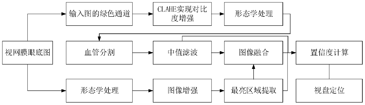
[0084] Step A: use image processing and sliding window methods to locate the optic disc area of ​​the retinal fundus image, and extract the located optic disc area as an area of ​​interest (ie ROI area) from the retinal fundus image to obtain the optic disc area image of the retinal fundus image, The process of locating the optic disc area is as follows image 3 shown.

[0085] Step A1. Morphologically process the original color retinal fundus image to obtain a grayscale enhanced retinal fundus image F':

[0086] Since different fundus imaging equipment under different environmental conditions will result in low fundus contrast or overall dark fundus, morphological processing can be used to...

Embodiment 2

[0153] For the original color retinal fundus image [such as Figure 7 (a)] (with a size of 2048×3047) to extract the cup-to-disk ratio. The first step is the positioning of the optic disc. First, the original color retinal fundus image is replaced with a grayscale image, and the bottom hat transformation is performed on the grayscale image to obtain the bottom hat transformed image [eg Figure 7 As shown in (b)], the same top-hat transformation is performed on the gray-scale fundus image to obtain the top-hat transformation image [such as Figure 7 (c)]. Then combine the obtained top-hat transformed image, bottom-hat transformed image and the original gray-scale fundus image to calculate the gray-scale retinal fundus enhanced image according to formula (1) [eg Figure 7 (d)]. Then extract the brightest area of ​​the grayscale retinal fundus enhanced image by extracting the histogram of the enhanced retinal fundus image, using iterative accumulation to select the brightest ...

Embodiment 3

[0158] Raw color retinal fundus images [eg Figure 9 (a)] (with a size of 2048×3047) to extract the cup-to-disk ratio. The first step is to position the optic disc. First, the original color retinal fundus image is replaced with a grayscale image, and the bottom hat transformation is performed on the grayscale image to obtain the bottom hat transformed image [eg Figure 9 Shown in (b)], the same top-hat transformation is performed on the gray-scale fundus image to obtain the top-hat transformation image [such as Figure 9 (c)]. Then combine the obtained top-hat transformed image, bottom-hat transformed image and the original gray-scale fundus image to calculate the gray-scale retinal fundus enhanced image according to formula (1) [eg Figure 9 (d)]. Then extract the brightest area of ​​the grayscale retinal fundus enhanced image by extracting the histogram of the enhanced retinal fundus image, using iterative accumulation to select the brightest pixel that accounts for 6.5...


PUM


Abstract

The invention discloses a retinal fundus image cup/disc ratio automatic evaluation method. The method comprises the following steps: A, extracting an optic disk area image from a retinal fundus image;B, establishing and training a optic disc optic cup segmentation network based on the deep convolutional neural network; C, acquiring a to-be-detected optic disc area image from the to-be-detected retina fundus image according to the step A, and inputting the to-be-detected optic disc area image into an optic disc optic cup segmentation network to output a to-be-detected optic disc segmentation mask image and a to-be-detected optic cup segmentation mask image; And step D, calculating the cup-disc ratio of the retinal fundus image according to the to-be-detected optic disc segmentation mask image and the to-be-detected optic cup segmentation mask image. The method is high in operation speed, good in effect, free of manual participation, low in cost and high in universality, and can be widely applied to auxiliary screening of glaucoma.

Description

technical field [0001] The invention belongs to the field of image information processing, in particular to a method for automatically evaluating the cup-to-disk ratio of retinal fundus images based on image processing and deep convolutional neural networks. Background technique [0002] Glaucoma is the second leading cause of blindness worldwide and the most serious cause of irreversible blindness. Although glaucoma cannot be completely cured, early detection and treatment can effectively reduce the possibility of blindness. Ophthalmologists often use Heidelberg Retinal Tomography (HRT) and Ocular Coherence Tomography (OCT) to detect glaucoma, but these procedures are time-consuming and require professional skills to operate the instruments. However, digital fundus images are widely used in glaucoma detection and are more economical and accurate. Compared with OCT and HRT, digital fundus cameras are easier to be used for basic ophthalmic disease diagnosis. In addition, t...

Claims


Application Information

Patent Timeline
IPC IPC(8): G06T7/00G06T7/11G06T7/12G06T5/00G06T5/30G06T5/50G06T7/62G06T7/80
Inventor 郭璠赵鑫谢斌赵于前廖胜辉梁毅雄邹北骥麦宇翔
Owner CENT SOUTH UNIV
Why Eureka
Try Eureka