CT image detection method and device based on deep learning and control equipment

A CT image and deep learning technology, applied in image analysis, image data processing, instruments, etc., can solve the problems that the precision is difficult to reach the expert level, the generalization ability is not strong, and it is difficult to apply clinically, so as to achieve fast response speed and ease Effects of medical pressure and difficulty in procrastinating illness

Pending Publication Date: 2019-05-31
清影医疗科技(深圳)有限公司
View PDF4 Cites 18 Cited by
  • Summary
  • Abstract
  • Description
  • Claims
  • Application Information

AI Technical Summary

Problems solved by technology

[0003] But so far there is no auxiliary diagnostic algorithm that can reach the clinical level, the main reason is that the accuracy of the algorithm is not high and the generalization ability is not strong.
For example, the level of the detection algorithm for liver cancer mainly depends on the results of the Liver TumorSegmentation Challenge. Among them, the fitting degree of liver segmentation can reach up to 0.96, but under the premise of low false alarm rate, the fitting degree of lesions can only reach 0.702 at most, and the precision Difficult to reach the level of experts, difficult to carry out clinical application

Method used


Image

  • CT image detection method and device based on deep learning and control equipment
  • CT image detection method and device based on deep learning and control equipment
  • CT image detection method and device based on deep learning and control equipment

Examples


Embodiment 1

[0041] This embodiment takes the CT image of liver cancer as an example for illustration, but it does not mean that the present invention can only be applied to the detection of liver cancer. The present invention can be widely used in the detection of CT images of different types of diseases. disease detection probability.

[0042] CT images reflect the human body's absorption coefficient of X-rays, but the density difference of each tissue structure, that is, the relative density, if a tissue has a lesion, its density will change, but because the comparison of the absorption coefficient is very cumbersome, the tissue and organ The absorption coefficient of X-rays is converted into CT value, and the unit is Hu. The CT value of water is 0Hu, and other different tissue densities are compared with it. The density greater than water is defined as a positive value, and the density smaller than water is defined as a negative value.

[0043] like figure 1 As shown, it is a flow cha...

Embodiment 2

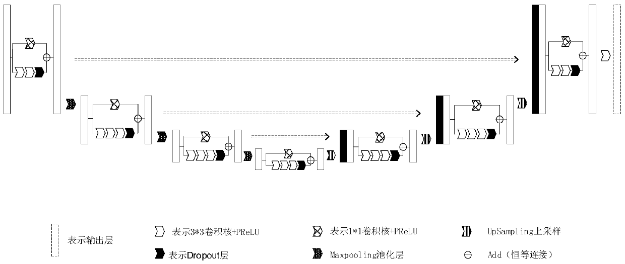
[0074] like Figure 8 As shown, it is a structural block diagram of the CT image detection device based on deep learning in this embodiment, including: a preprocessing module: used to preprocess the original image to obtain an image to be detected; an image detection model acquisition module: used to build a neural network detection model, and perform model training to obtain a neural network CT image detection model; output detection result module: used to input the image to be detected into the neural network CT image detection model to obtain detection results.

[0075] On the other hand, the present invention also includes a deep learning-based CT image detection control device, including: at least one processor; and a memory connected to at least one processor; wherein, the memory stores information that can be processed by at least one The instructions executed by the processor are executed by at least one processor, so that the at least one processor can execute the met...


PUM


Abstract

The invention discloses a CT image detection method based on deep learning. The method comprises the steps of preprocessing an original image to obtain a to-be-detected image; establishing a trainingneural network detection model to obtain a neural network CT image detection model; inputting the to-be-detected image into the neural network CT image detection model; Obtaining detection results, wherein the input to-be-detected image comprises cross section direction information. Different from an existing single-input and single-output model training mode, input is changed into five-dimensional data from a one-dimensional data image of a single slice, output is also changed into two-channel mask output from single output, model precision and focus fitting degree of a prediction result areimproved, precision reaches expert level, and clinical application can be carried out. Moreover, in the process, a neural network CT image detection model is established by using big data in advance,during judgment, a CT scanning image of a patient is directly input, the model is used for calculation, the model output response speed is high, and disease delay is not likely to be caused.

Description

technical field [0001] The present invention relates to the field of image detection, in particular to a CT image detection method, device and control equipment based on deep learning. Background technique [0002] Cancer is one of the most common causes of death due to disease in the world, and the number of people who die from cancer remains high every year. Due to the serious threat cancer poses to human beings, people have made many efforts to carry out early diagnosis and treatment. With the development of imaging technology, three-dimensional CT (computerized tomography) has become a routine clinical method for cancer diagnosis. With the emergence of high-resolution CT scanners in recent years, more and more CT image data are generated. Surgeons and radiologists often need to manually label thousands of CT scan slice images, which is a heavy burden for them. Therefore, efficient medical aided diagnosis algorithms have become an urgent need. [0003] But so far, no aux...

Claims


Application Information

Patent Timeline
Patent Type & Authority Applications(China)
IPC IPC(8): G06T7/00
Inventor 邹昊郭玉成谢苏李晓禹
Owner 清影医疗科技(深圳)有限公司
Why Eureka
Try Eureka