mip3α-fgfr1-pd1/fc fusion protein and its nucleic acid molecule and application
A -FGFR1-PD1, FGFR1 technology, applied in the field of anti-tumor drugs, can solve the problems of difficult specification, complicated in vitro operation, high cost, and achieve the effect of inhibiting growth, reducing tumor volume and reducing tumor mass
- Summary
- Abstract
- Description
- Claims
- Application Information
AI Technical Summary
Problems solved by technology
Method used
Image
Examples
preparation example Construction
[0047] The present invention also provides a method for preparing nucleic acid molecules described in the above technical solution, comprising the following steps:
[0048] 1) Mix primers 1 to 84, and use primers 1 to 84 as templates and primers to perform the first round of PCR amplification to obtain a mixed amplification product;
[0049] The nucleotide sequences of the primers 1 to 84 are sequentially shown as SEQ ID NO.3 to SEQ ID NO.86;
[0050] 2) Using the mixed amplification product as a template, primer 1 as an upstream primer, and primer 84 as a downstream primer, perform a second round of PCR amplification to obtain a nucleic acid molecule of the MIP3α-FGFR1-PD1 / Fc fusion protein.
[0051] In the present invention, the nucleotide sequences of the primers 1 to 84 are preferably shown in SEQ ID NO.3 to SEQ ID NO.86.
[0052] In the present invention, the primers 1 to 84 are preferably artificially synthesized, and equal amounts of the obtained primers 1 to 84 are mi...
Embodiment 1M
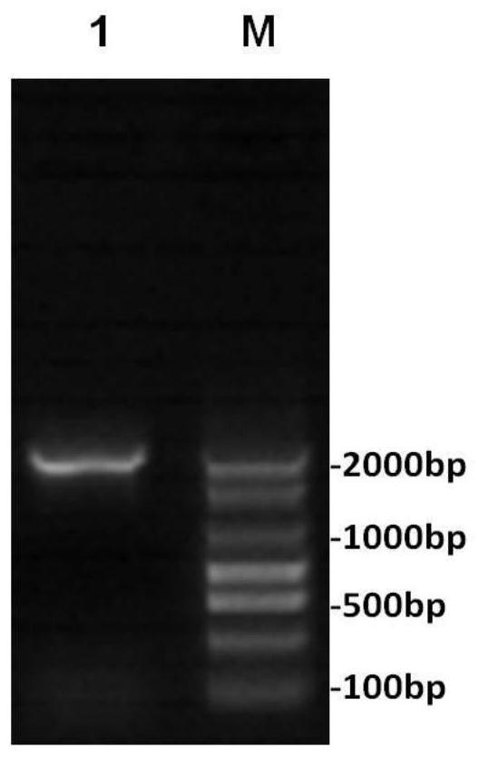
[0073] Example 1 Construction of Nucleic Acid Molecules of MIP3α-FGFR1-PD1 / Fc Fusion Protein
[0074] 1) Mix primers 1 to 84 in equal amounts to obtain a primer mixture with a concentration of 20 mM for each of primers 1 to 84. Primers 1 to 84 in the primer mixture serve as primers and templates for each other, and perform the first round of PCR amplification according to the following conditions: to obtain mixed amplification products.
[0075] The PCR amplification system used in the first round of PCR amplification is 50 μL: 33 μL ddH 2 O. 5 μL of 10×PCRBuffer, 1 μL of dNTP with a concentration of 2.5 mM, 10 μL of a mixture of primers 1 to 84 with a concentration of 20 mM, and 1 μL of Pfu high-fidelity DNA polymerase with a concentration of 2.5 U / L;
[0076] The PCR reaction program used in the first round of PCR amplification is: 95°C for 5 minutes; 94°C for 30s, 55°C for 30s, 72°C for 30s, cycle 25 times; 72°C for 5 minutes.
[0077] 2) Using the mixed amplification pro...
Embodiment 2
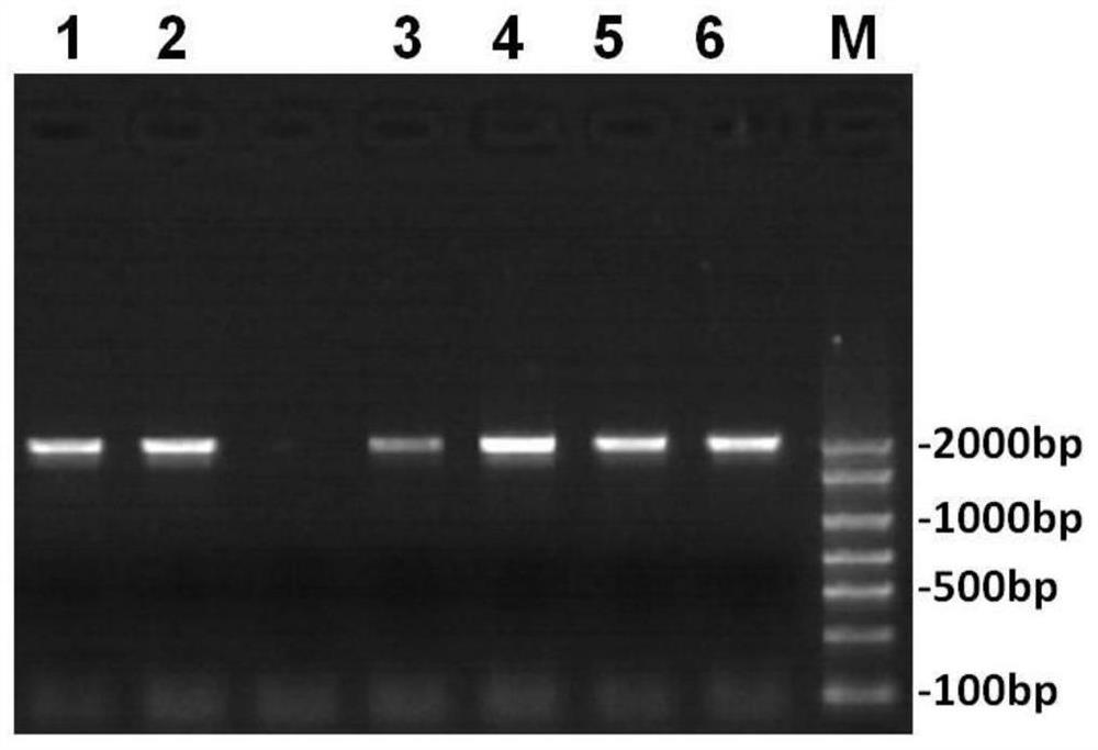
[0096] Construction and screening, identification of embodiment 2 target plasmids
[0097] 1) Preparation of LB medium
[0098] Liquid medium: tryptone 10g, yeast extract 5g, NaCl 10g, add 950ml of deionized water to dissolve, adjust to pH 7.2 with 5mol / L NaOH, finally add water to 1000ml, aliquot and autoclave at 10 pounds for 20min.
[0099] Solid medium: 1.5g of agar powder, dissolved in 100ml of LB liquid medium, sterilized under 10 pounds of high pressure for 20min.
[0100] 2) Digestion of target gene fragments and plasmids
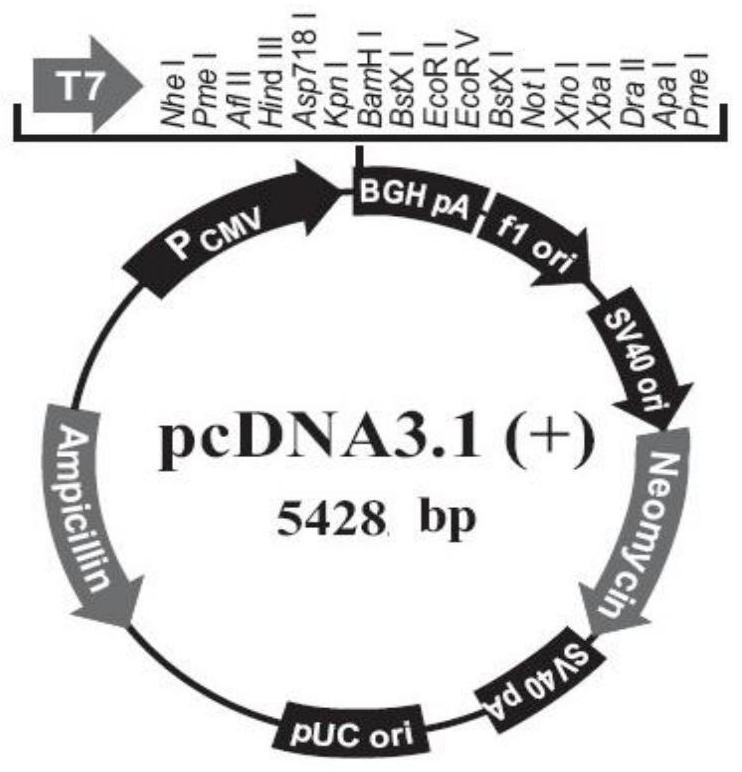
[0101] Carry out double digestion of pcDNA3.1(+) with AflII and XbaI, the enzyme digestion reaction system is: pcDNA3.1(+) 12μL, 10buffer 2μL, AflII1μl, XbaI 1μL and ddH 2 O 4 μL, a total of 20 μL.
[0102] Put the above system in a water bath at 37°C for 6h, then inactivate the restriction enzyme at 65°C for 15min. The digested products were recovered and purified by gel recovery kits from Jiangsu Futai Biotechnology Co., Ltd. respectively. Aft...
PUM
| Property | Measurement | Unit |
|---|---|---|
| molecular weight | aaaaa | aaaaa |
| molecular weight | aaaaa | aaaaa |
| purity | aaaaa | aaaaa |
Abstract
Description
Claims
Application Information
 - R&D
- Intellectual Property
- Life Sciences
- Materials
- Tech Scout
- Unparalleled Data Quality
- Higher Quality Content
- 60% Fewer Hallucinations
Browse by: Latest US Patents, China's latest patents, Technical Efficacy Thesaurus, Application Domain, Technology Topic, Popular Technical Reports.
© 2025 PatSnap. All rights reserved.Legal|Privacy policy|Modern Slavery Act Transparency Statement|Sitemap|About US| Contact US: help@patsnap.com


