Thyroid nodule ultrasonic image segmentation method based on deep learning

A technology for thyroid nodules and ultrasound images, applied in neural learning methods, image analysis, image enhancement, etc., can solve problems such as low accuracy, low resolution, and difficult segmentation, and achieve the effect of improving work efficiency and accuracy

Pending Publication Date: 2019-07-26
TIANJIN UNIV
View PDF5 Cites 26 Cited by
  • Summary
  • Abstract
  • Description
  • Claims
  • Application Information

AI Technical Summary

Problems solved by technology

[0008] The invention provides a method for segmenting ultrasound images of thyroid nodules based on deep learning. The invention effectively overcomes the problems of difficult segmentation and low accuracy caused by low resolution of ultrasound images of thyroid nodules and a lot of interference information, and finally greatly improves the For the accuracy of nodule segmentation, see the description below:

Method used


Image

  • Thyroid nodule ultrasonic image segmentation method based on deep learning
  • Thyroid nodule ultrasonic image segmentation method based on deep learning
  • Thyroid nodule ultrasonic image segmentation method based on deep learning

Examples


Embodiment 1

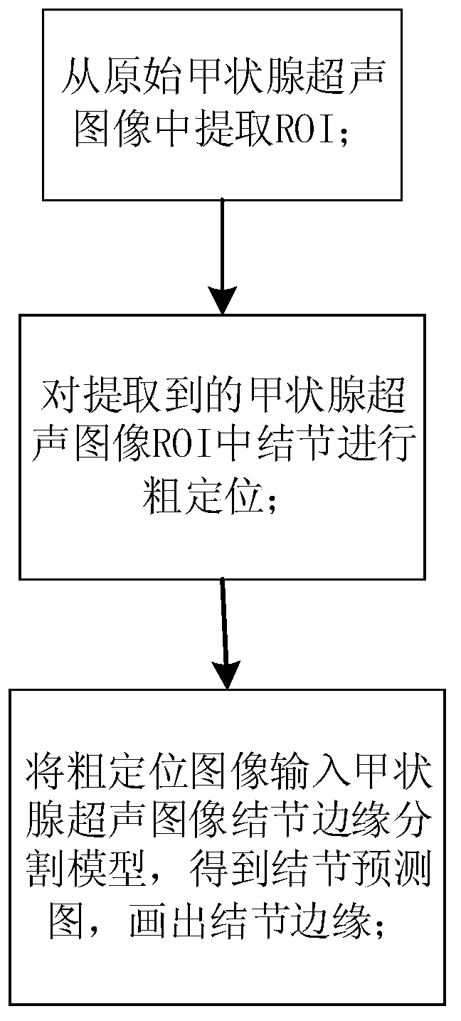
[0029] In order to solve the problems existing in the background technology, the embodiment of the present invention proposes a deep learning-based ultrasound image segmentation method for thyroid nodules, see figure 1 , the method includes the following steps:

[0030] 101: extract a region of interest (Region of Interest, ROI) from the original thyroid ultrasound image;

[0031] Among them, the ultrasound image can be divided into ROI and background area, where ROI is the area containing the echo information of the internal shape and structure of the thyroid gland, and the background area is other parts except ROI, including: ultrasound scanner model, various scanning parameters and scanning parameters. Information such as location that has nothing to do with the thyroid itself.

[0032] 102: Input the extracted thyroid ultrasound image ROI, and roughly locate the nodules in the ROI;

[0033] 103: Input the rough positioning image into the nodule edge segmentation model of...

Embodiment 2

[0044] Combine below figure 1 1. The specific calculation formula further introduces the scheme in Embodiment 1, see the following description for details:

[0045] 201: In the process of segmenting an ultrasound image of a thyroid nodule, ROI must first be extracted from the original ultrasound image of a thyroid nodule;

[0046] An embodiment of the present invention proposes a fully convolutional neural network model to perform ROI semantic segmentation on thyroid ultrasound images. In order to learn the global information of the image as much as possible, the fully convolutional neural network structure uses 10 convolutions and 5 pooling to ensure that the receptive field of the final pixel classification result contains the entire image as much as possible.

[0047] 202: The ROI segmentation model selects cross entropy as a loss function, as shown in formula (1).

[0048]

[0049] Among them, y i is the true label of category i, p i Is the probability value of cate...

Embodiment 3

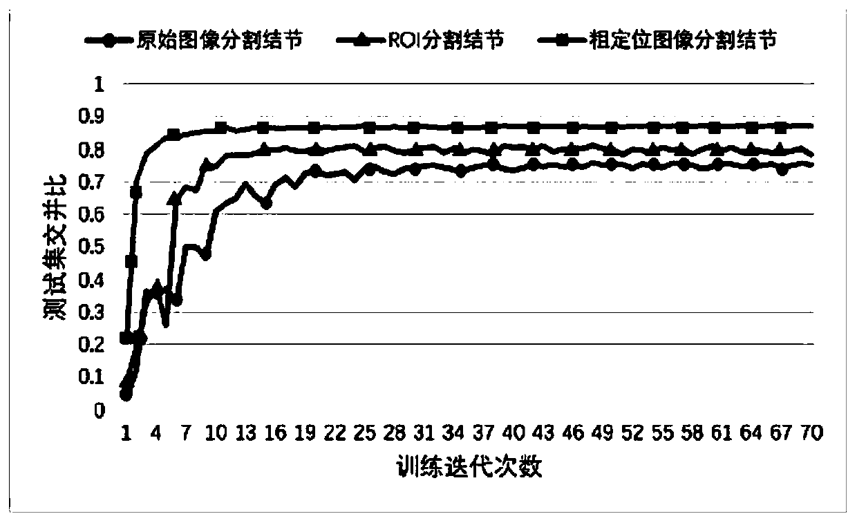
[0063] Below in conjunction with specific calculation formula, example, the scheme in embodiment 1 and 2 is carried out feasibility verification, see the following description for details:

[0064] In order to compare the influence of different segmentation levels on the segmentation results, 1000 original ultrasound image datasets provided by the Tianjin Medical University Cancer Hospital were used as the input original images, and the nodule edge annotations under the guidance of radiologists under the original images of thyroid ultrasound images were used as the original images. Labeled images, 80% as training set and 20% as testing set, were segmented using a thyroid ultrasound image nodule edge segmentation model. At the same time, the original ultrasound image of the thyroid gland was segmented into ROI, and the label image after labeling the edge of the nodule was segmented synchronously under the guidance of a radiologist to obtain a nodule segmentation dataset under th...


PUM


Abstract

The invention discloses a thyroid nodule ultrasonic image segmentation method based on deep learning, and the method comprises the following steps: inputting an original thyroid ultrasonic image, obtaining an interested region prediction image through an interested region semantic segmentation model, and enabling the prediction image to correspond to the original thyroid ultrasonic image, and extracting an interested region; inputting the extracted thyroid ultrasound image region of interest into the artificial mark recognition model, and roughly positioning nodules in the region of interest;inputting the coarse positioning image into a thyroid ultrasound image nodule edge segmentation model to obtain a nodule prediction image, and drawing a nodule edge. According to the method, the problems of segmentation difficulty, low accuracy and the like caused by low resolution and much interference information of the thyroid nodule ultrasonic image are effectively solved, and finally the nodule segmentation accuracy is greatly improved.

Description

technical field [0001] The present invention relates to the field of ultrasonic image segmentation, in particular to a method for segmenting ultrasonic images of thyroid nodules based on deep learning. Background technique [0002] Image semantic segmentation is to divide the image into different regions according to different semantics, and visually mark the categories of objects represented by these regions through different colors. Medical image segmentation is one of the hot applications in the field of image semantic segmentation. The purpose of medical image segmentation is to divide the image into different parts according to the semantic characteristics of the image itself, so as to provide a reliable basis for clinical diagnosis and pathological research. The accuracy of the segmentation not only affects other processing steps, but also may affect the image quality. The analysis results will have an impact on clinical diagnosis and scientific research. [0003] Th...

Claims


Application Information

Patent Timeline
Patent Type & Authority Applications(China)
IPC IPC(8): G06T7/00G06T7/11G06T7/12G06N3/04G06N3/08
CPCG06T7/0012G06T7/11G06T7/12G06N3/08G06T2207/10132G06T2207/20081G06T2207/20084G06T2207/30096G06T2207/20104G06N3/045
Inventor 高洁高明牛文倩徐天一赵满坤魏玺张晟王臣汉刘志强于瑞国喻梅尉智辉
Owner TIANJIN UNIV
Why Eureka
Try Eureka