
Vaginal secretion detection device and method

A technology for vaginal secretions and a detection device, applied in the fields of biological and medical detection, can solve the problems of error-prone counting and input, complicated device, low detection rate of the instrument, etc., so as to improve the detection speed and recognition accuracy, and simplify the complexity of the instrument. , the effect of improving the detection efficiency

Pending Publication Date: 2019-08-16
瓜藤生物科技(深圳)有限公司
View PDF0 Cites 7 Cited by
  • Summary
  • Abstract
  • Description
  • Claims
  • Application Information

AI Technical Summary

Problems solved by technology

The method of manual microscopic examination not only requires manual smears, constantly switching the field of view to count, but also corresponding the results of microscopic examination with the results of dry chemical testing, and manual input, which is time-consuming and laborious, and the detection efficiency is low, and the counting and input process is prone to errors
[0004] At present, for microscope inspection, there are also automatic detection devices on the market, but the devices are complicated, the recognition rate of formed components is low, and it takes a long time
Moreover, the pictures taken are stored locally, and the detection and identification of the formed components of the sample is limited to the local area, without effective connection with the cloud platform
The low detection rate of instruments on the market is mainly due to the traditional classification method used. The traditional neural network classification method not only requires a lot of pre-processing of pictures, but also needs to artificially select some features when constructing and training the neural network. The selected features are not necessarily suitable for computer recognition, and are prone to overfitting
Moreover, since the detection of formed components is limited to the local area, the large number of samples cannot correct the detection model in a timely and effective manner, and the detection accuracy will not increase with the increase of detection samples.

Method used

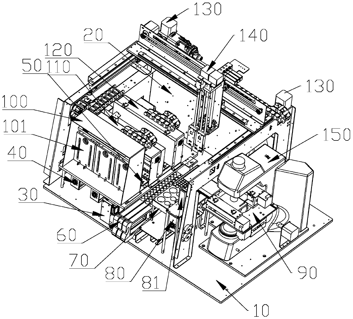
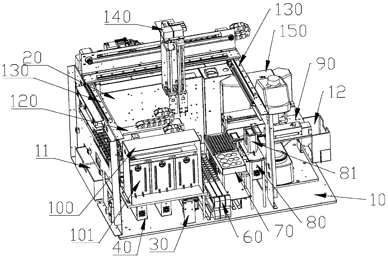
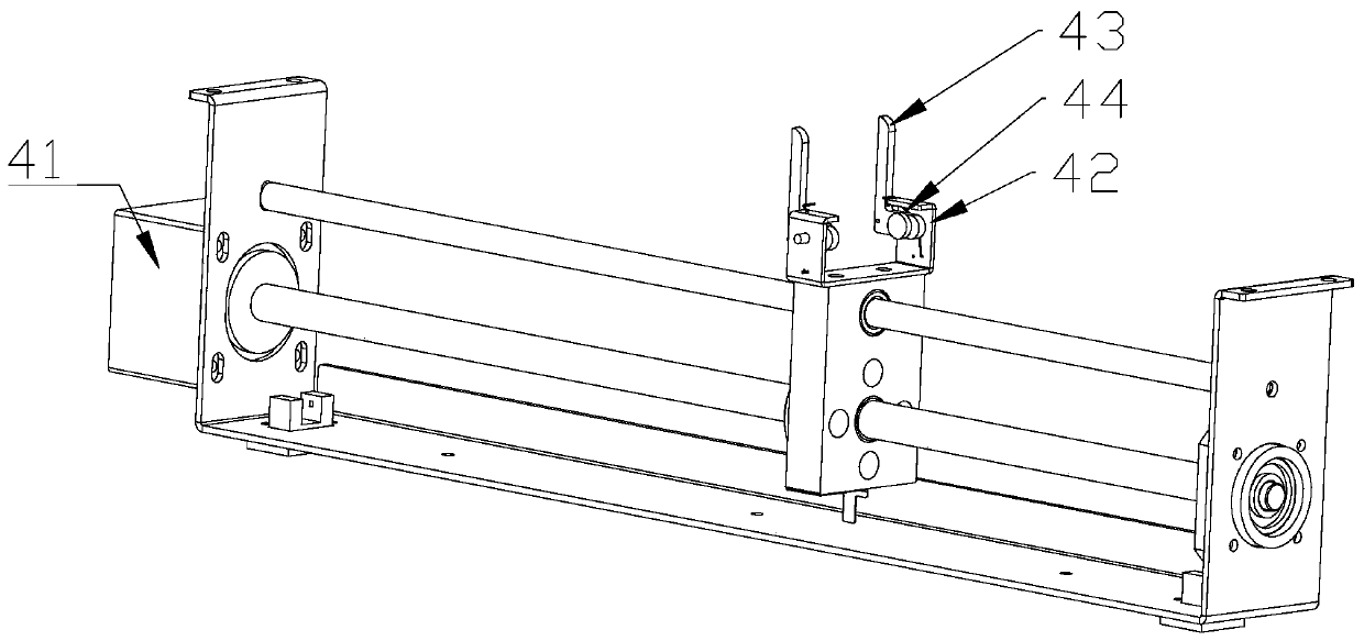

Image

  • Vaginal secretion detection device and method
  • Vaginal secretion detection device and method
  • Vaginal secretion detection device and method

Examples


Embodiment Construction

[0040] It should be noted that, in the case of no conflict, the embodiments in the present application and the features in the embodiments can be combined with each other. The present invention will be further described in detail below in conjunction with the drawings and specific embodiments.

[0041] If there are directional indications (such as up, down, left, right, front, back...) in the embodiments of the present invention, they are only used to explain the relative positions of the components in a certain posture (as shown in the drawings) relationship, motion, etc., if the particular pose changes, the directional indication changes accordingly.

[0042] In addition, in the present invention, the descriptions involving "first", "second" and so on are only for the purpose of description, and should not be understood as indicating or implying their relative importance or implicitly indicating the quantity of the indicated technical features. Thus, the features defined as ...


PUM


Abstract

Embodiments of the invention disclose a vaginal secretion detection device and method. The detection device comprises a base and a control circuit board; a bar code scanning head, an upper card assembly and an incubation assembly are sequentially arranged on one side of the base; the other side of the base is sequentially provided with a test tube rack assembly, a consumable replacement assembly,a slide upward and downward moving assembly and a microscope electric platform; the upper card assembly is provided with a card storage box; a reagent card clip is arranged in the card storage box; afirst optical detection assembly and a second optical detection assembly are arranged above the incubation assembly; a reagent card recovery box is arranged on the outer side of the incubation assembly; a Y-direction driving assembly is further arranged on the base; a liquid shifting assembly is arranged on the Y-direction driving assembly; a microscope is arranged above the microscope electric platform; a slide recovery box is arranged on the outer side of the microscope electric platform; and a slide clip is arranged on the slide upward and downward moving assembly. According to the vaginalsecretion detection device and method, the detection speed and the recognition accuracy are improved; and manual feature extraction and image preprocessing are not needed, so that the misdiagnosis isreduced.

Description

technical field [0001] The invention relates to the technical field of biological and medical detection, in particular to a vaginal secretion detection device and method. Background technique [0002] The detection of vaginal secretions is generally divided into two parts, dry chemical detection and microscopic examination. Dry chemical detection is a semi-quantitative detection. Generally, when the dry chemical test results show that the patient's physical indicators are abnormal, a microscopic examination is performed on the patient to complete the confirmation of the test results. However, the current microscopic examination of vaginal secretions is mainly through manual detection. [0003] After the tester gets the patient sample, he rinses the sample. After rinsing, he makes a manual smear and puts it under the microscope for testing. The formed components in the film are classified and counted. Formed components mainly include: white blood cells, Candida, clue cell...

Claims


Application Information

Patent Timeline
Patent Type & Authority Applications(China)
IPC IPC(8): G01N35/00G01N35/10G01N21/84G06T7/00
CPCG01N35/00584G01N35/00732G01N35/1011G01N21/84G01N21/8483G06T7/0012G01N2035/00752G01N2035/00465G06T2207/10004G06T2207/10056G06T2207/20081G06T2207/20084G06T2207/30004
Inventor 黄炜中王振刚吴涛梁桂彬赵明建刘波李静
Owner 瓜藤生物科技(深圳)有限公司
Learn More