Medical image preprocessing method

A medical image and preprocessing technology, which is applied in the field of medical image processing, can solve problems such as inability to effectively process ultra-high resolution digital medical images, different area ratios, difficult balance between calculation amount and accuracy, and achieve efficient and rapid prediction. The effect of processing, improving efficiency, and improving loading efficiency

Active Publication Date: 2019-09-17
SUN YAT SEN UNIV
View PDF3 Cites 6 Cited by
  • Summary
  • Abstract
  • Description
  • Claims
  • Application Information

AI Technical Summary

Problems solved by technology

[0005] The existing classification and recognition technology based on image segmentation is generally used for pathological image analysis at small resolutions, and it is still unable to effectively deal with such a huge amount of data in ultra-high-resolution digital medical images
In addition, in medical image recognition tasks, on the one hand, the samples used include postoperative large slice samples and puncture samples used in early screening; on the other hand, the shape and area ratio of tissues in each digital slice are different. , making the amount of calculation and accuracy of sample extraction a pair of contradictions that are difficult to balance

Method used


Image

  • Medical image preprocessing method
  • Medical image preprocessing method
  • Medical image preprocessing method

Examples


Embodiment 1

[0063] This embodiment provides the medical image preprocessing method of the present invention, which includes the following steps:

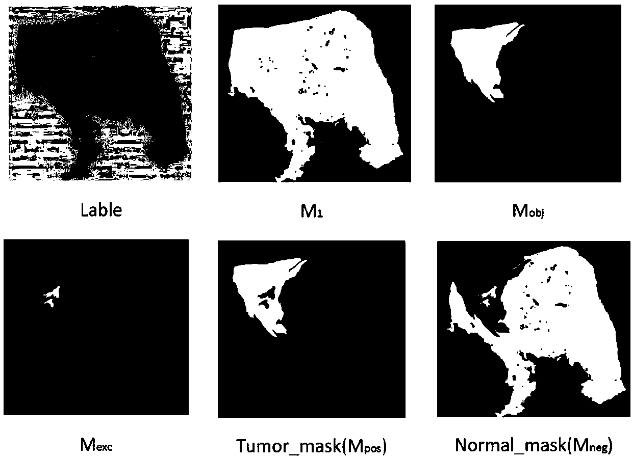
[0064] Semantic imaging of label information: pre-read digital medical images and their label information, and apply discriminant algorithms to convert formatted text label information into multi-level classification mask images;

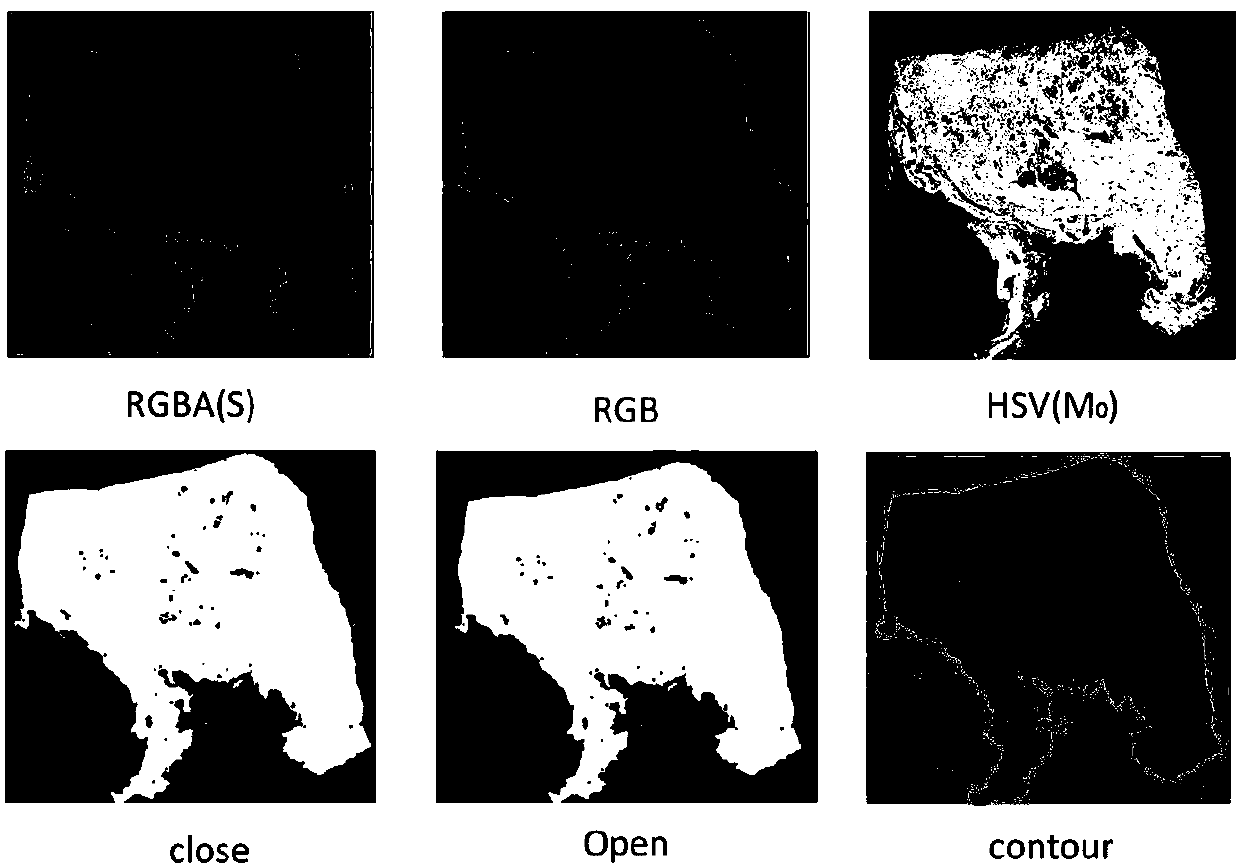
[0065] Region of Interest Extraction: Read digital medical images, remove the transparency channel to acquire images, extract the outline of the tissue region in the current slice image, and divide the image into tissue regions and background regions, such as figure 1 shown;

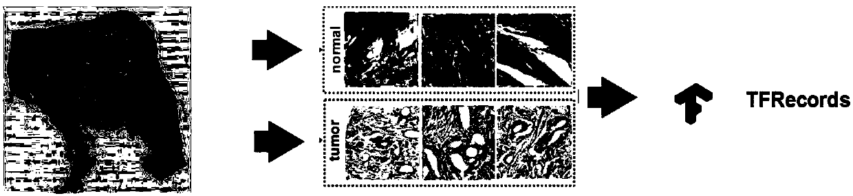
[0066] Multi-mask sample classification extraction: use the generated multi-level classification mask to extract positive samples and negative samples in the tissue area, such as figure 2 with image 3 shown; and encapsulate the sample data information to form structured data that can be applied to neural network model training and predictio...

Embodiment 2

[0069] This embodiment is a preferred implementation on the basis of the above-mentioned embodiment 1. The difference between this embodiment 2 and the above-mentioned embodiment is that: in this embodiment, the step of label information semantic imaging has the following: One or more preferred implementations, these implementations can be implemented individually or in combination:

[0070] In some implementations, the transparency channel is an Alpha channel.

[0071] In some implementations, the format of the text tag is XML format. The format of the text label may adopt multiple formats. In one implementation manner, the format is XML format, and other corresponding different formats may be adopted in other implementation manners according to actual needs.

[0072] In some implementations, the discriminant algorithm is a closed polygon coordinate discriminant algorithm. In this embodiment, the discriminant algorithm adopts a closed polygon coordinate discriminant algorit...

Embodiment 3

[0092] This embodiment is a preferred implementation based on the above-mentioned embodiment 1. The difference between this embodiment 3 and the above-mentioned embodiment is that in this embodiment, the region of interest extraction step has one of the following Or multiple preferred implementations, these implementations can be implemented alone or in combination:

[0093] In some embodiments, the method of reading the digital medical image is reading using Openslide.

[0094] In some embodiments, the image is an RGB image. The images in this technical solution may be RGB images, but may also be images in other formats, depending on the specific implementation.

[0095] In some implementations, after the image is acquired, it further includes extracting the outline of the tissue region by using color gamut space shift, erosion and dilation. Through this technical feature, the tissue area in the medical image can be quickly defined by the preset color threshold, and further...


PUM


Abstract

The invention discloses a medical image preprocessing method which comprises the following steps of pattering a label information meaning, pre-reading a digital medical image and label information thereof, and applying a discriminating algorithm for converting text label information with a format to a multilayer classified mask image; extracting an interested area, namely reading the digital medical image, eliminating a transparency channel acquisition image, extracting a tissue area contour in a current slice image, and dividing the image to a tissue area and a background area; and performing classified extraction of multiple mask samples, namely extracting a positive sample and a negative sample in the tissue area by means of the generated multilayer classified mask, and packaging the sample data information, thereby forming structured data which can be applied to neural network model training and predicting. Through the medical image preprocessing method, data preprocessing with higher efficiency and higher accuracy can be realized.

Description

technical field [0001] The invention relates to the technical field of medical image processing, in particular to a method for preprocessing medical images. Background technique [0002] The following content is only the background introduction of the technology of the present application as recognized by the inventor, and does not necessarily constitute the prior art. [0003] Computer aided diagnosis (computer aided diagnosis, CAD) refers to the method of assisting in the detection of lesions and improving the accuracy of diagnosis through imaging, medical image processing technology and other possible physiological and biochemical means, combined with computer analysis and calculation. As the "third eye" of doctors, the wide application of CAD system helps to improve the sensitivity and specificity of doctors' diagnosis. [0004] In order to use this information accurately and efficiently, computer-aided diagnosis research based on cancer medical images has become a hot ...

Claims


Application Information

Patent Timeline
Patent Type & Authority Applications(China)
IPC IPC(8): G16H30/20
CPCG16H30/20
Inventor 王国利李亮郭斌刘力徐琰
Owner SUN YAT SEN UNIV
Why Eureka
Try Eureka