Endoscopic lens unit

An endoscope and lens technology, applied in the field of medical lenses, can solve the problems of depth affecting lens observation, inconvenient inspection, small field of view, etc., to improve edge resolution capability, increase range and depth, and reduce lens aperture. Effect

Active Publication Date: 2019-09-20
合肥联创光学有限公司
View PDF6 Cites 2 Cited by
  • Summary
  • Abstract
  • Description
  • Claims
  • Application Information

AI Technical Summary

Problems solved by technology

At present, most endoscopes have large calibers, and the process of entering the human body is more painful, and it is not convenient to inspect the lesion with a narrow channel.
[0003] However, the current endoscope lens still has the problems of large caliber, small depth of field, and small field of view. The smaller the caliber of the endoscope, the less discomfort it will cause to the human body, and the large caliber of the endoscope will increase the production cost. At the same time, the field of view and depth of field are also important parameters for the use of endoscopes. A small field of view will affect the scope of lens observation, while a small depth of field will affect the depth of lens observation.

Method used


Image

  • Endoscopic lens unit
  • Endoscopic lens unit
  • Endoscopic lens unit

Examples


Embodiment 1

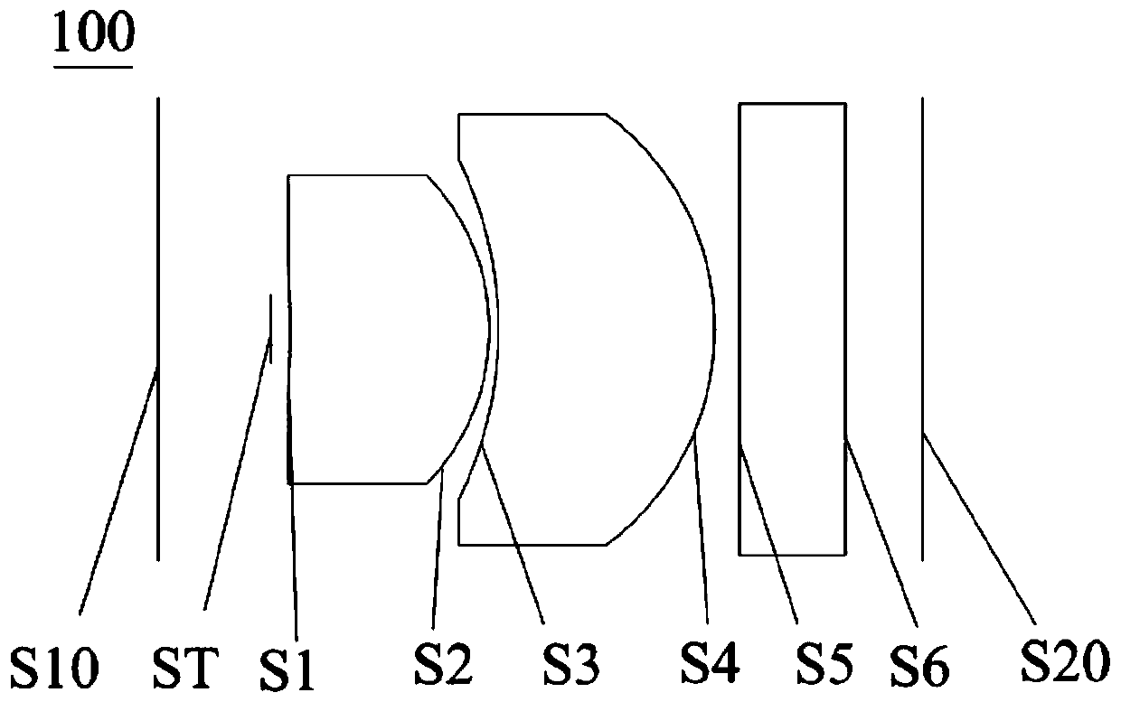
[0081] See figure 1 The endoscope lens 100 provided by the first embodiment of the present invention includes a first lens L1, a second lens L2, a stop ST, and a filter G1 in order from the object plane S10 to the image plane S20.

[0082] The first lens L1 has a positive refractive power, the object side surface S1 is a concave surface, the image side surface S2 is a convex surface, and the first lens L1 is a glass aspheric lens.

[0083] The second lens L2 has a positive refractive power, the object side surface S3 is a concave surface, the image side surface S4 is a convex surface, and the second lens L2 is a glass spherical lens. In other embodiments of the present invention, the second lens L2 may be a plastic aspheric lens.

[0084] The stop ST is arranged in front of the first lens L1 (that is, between the first lens L1 and the object plane S10), and the filter G1 is arranged between the second lens L2 and the image plane S20. The object side of the filter G1 is S5, and the ...

Embodiment 2

[0093] See Figure 5 , The endoscope lens 200 provided by the second embodiment of the present invention. The endoscope lens 200 in this embodiment is substantially the same as the endoscope lens 100 in the first embodiment, except that the second lens L2 of the endoscope lens 200 in this embodiment has a negative focal length. For a high-degree plastic aspheric lens, the object side S3 is convex, and the image side S4 is concave. The curvature radius and material selection of each lens are different. For specific parameters of each lens, see Table 2-1.

[0094] table 2-1

[0095]

[0096]

[0097] The parameters of the aspheric surface of each lens in this embodiment are shown in Table 2-2:

[0098] Table 2-2

[0099]

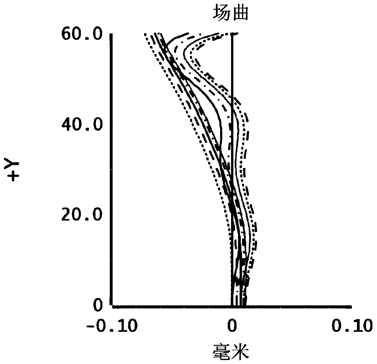
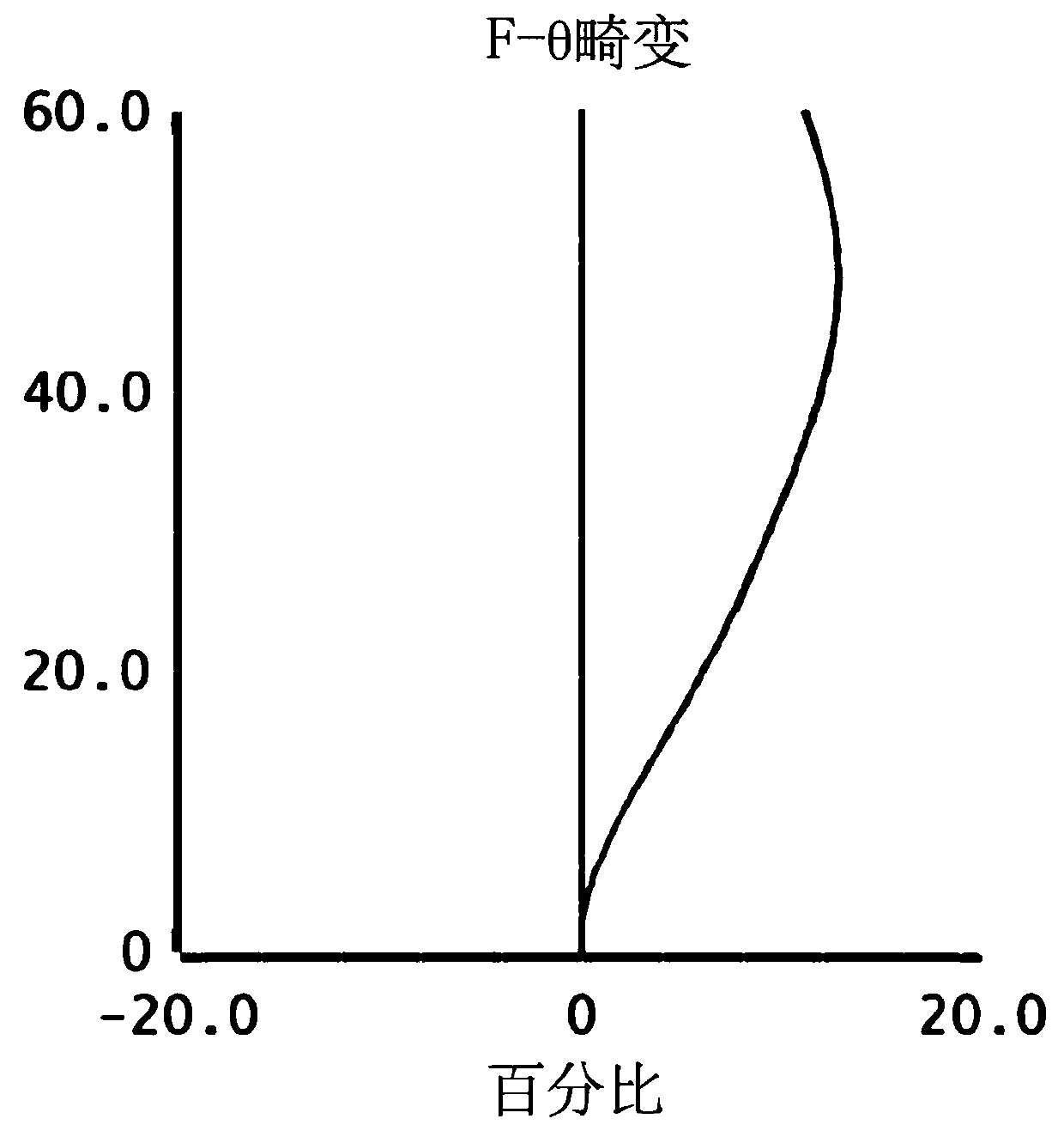
[0100] In this embodiment, the curvature of field, distortion, and axial chromatic aberration are as follows: Image 6 , Figure 7 with Figure 8 Shown. Since the smaller the data range of the image point, the better the performance of the lens. Figure 6 to Figure ...

Embodiment 3

[0102] See Picture 9 , The endoscope lens 300 provided by the third embodiment of the present invention. The endoscope lens 300 in this embodiment is substantially the same as the endoscope lens 100 in the first embodiment, except that the second lens L2 object side surface S3 of the endoscope lens 300 in this embodiment is convex. , The image side surface S4 is concave, and the radius of curvature and material selection of each lens are different. The specific parameters of each lens are shown in Table 3-1:

[0103] Table 3-1

[0104]

[0105]

[0106] The parameters of the aspheric surface of each lens in this embodiment are shown in Table 3-2:

[0107] Table 3-2

[0108]

[0109] In this embodiment, the curvature of field, distortion, and axial chromatic aberration are as follows: Picture 10 , Picture 11 with Picture 12 Shown. Since the smaller the data range of the image point, the better the performance of the lens. Figure 10 to Figure 12 It can be seen that curvature of f...


PUM


Abstract

The invention discloses an endoscopic lens unit, comprising, from the objective side to the image side, a first lens having a concave objective face and a convex image face; a second lens having positive or negative focal powder; and an optical filter arranged between the second lens and the image side, wherein the first lens is a glass or plastic non-spherical lens, and the second lens is a glass or plastic non-spherical lens. The endoscopic lens unit also comprises a diaphragm which is positioned between the first lens and the objective side, or between the first lens and the second lens. The endoscopic lens unit provided herein has the advantage that as the lenses, the optical filter and the diaphragm are reasonably matched and limited, a large visual angle and a large depth of field can be guaranteed at the premise of lens diameter reduced, observation range is widened, and observation depth is increased.

Description

Technical field [0001] The invention relates to the technical field of medical lenses, in particular to an endoscope lens. Background technique [0002] An endoscope is a medical device that penetrates into the human body through the natural pores of the human body, and observes the internal structure or pathological conditions through imaging images. At present, most endoscopes have large calibers, and the process of entering the human body is painful, and it is not convenient to check the lesions with narrow passages. [0003] However, the current endoscope lens still has the problems of large diameter, small depth of field, and small field of view. The smaller the diameter of the endoscope, the less discomfort caused to the human body, and the excessive diameter of the endoscope will increase the production cost. At the same time, the size of the field of view and the depth of field are also important parameters used by the endoscope. A small field of view will affect the range...

Claims


Application Information

Patent Timeline
Patent Type & Authority Applications(China)
IPC IPC(8): A61B1/00G02B23/24
CPCA61B1/00186A61B1/00071G02B23/243G02B13/003G02B9/04A61B1/00096A61B1/05
Inventor 魏文哲刘绪明曾吉勇王卓
Owner 合肥联创光学有限公司
Why Eureka
Try Eureka