Cancer cell recognition method, device and system
A recognition method and a technology of a recognition device, which are applied in the medical field, can solve problems such as low recognition rate, out-of-control division, and difficulty in curing malignant tumors, and achieve the effect of reducing background noise and difficulty
- Summary
- Abstract
- Description
- Claims
- Application Information
AI Technical Summary
Problems solved by technology
Method used
Image
Examples
Embodiment 1
[0032] With the development of global high-tech, smart medical care and safe treatment have become the focus of every country. One of the key points in the infrastructure required to realize this concept is that the ubiquitous artificial intelligence network will likely be embedded in Every medical device, including positron-emission CT, MRI, and electron microscopy systems, is then used to detect abnormal cells in the pictures. By detecting various forms of cells to distinguish cell types, the detected targets are simple cell tissues, for example, there are few cell types, and there is not much cell overlap, but in the face of more cell types, cell overlap is not easy to judge When it comes to cell tissue, the recognition rate is not high. In order to avoid misjudgment and accidents, we not only need to detect the size of the cells, but also predict the shape of the cells and the distribution density in the tissue. When the doctor gets the picture, he can see more prompt info...
Embodiment 2
[0051] The purpose of this embodiment is to further illustrate the principles / steps of the pre-order training work involved in the embodiment and the subsequent actual application process.
[0052] Establishment and training of recognition network for identifying cancer cells, including:
[0053] S01. Manually process the microscope video of the tissue section frame by frame, manually calibrate the area where cells appear in each picture, and obtain a calibration map for each cell, and each pixel in the calibration map can only be 0 or 1 ( 0 means that the pixel does not belong to the cell, and 1 means that the pixel belongs to the cell), the video image file number of the same tissue slice sample is V, and the number of the cell is N, then the corresponding calibration map is named V_N, and finally Obtain the calibration image set Ti for the image pi, that is, establish a training sample for a frame of image
[0054] S02, repeat S01, calibrate M-frame images, for example, M>...
Embodiment 3
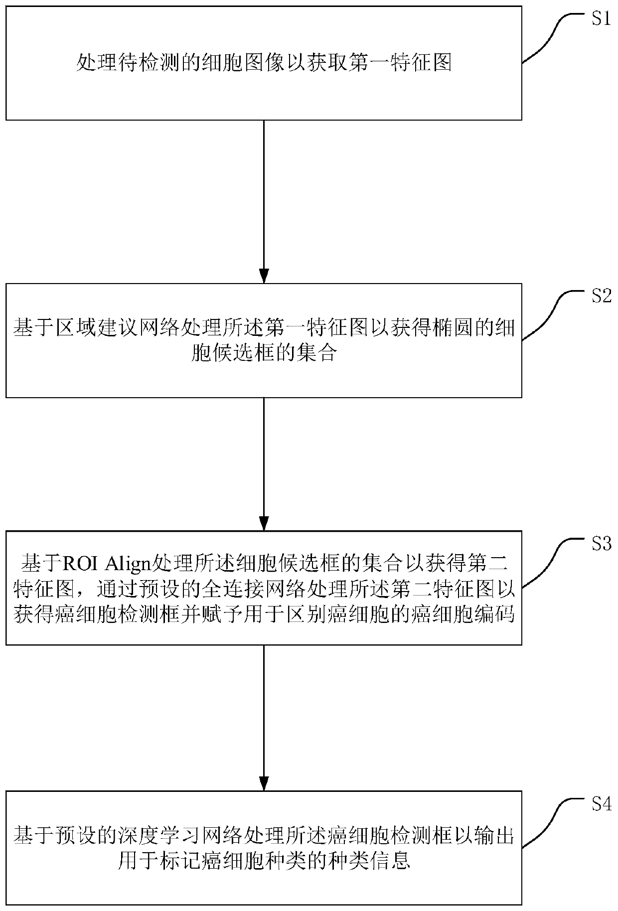
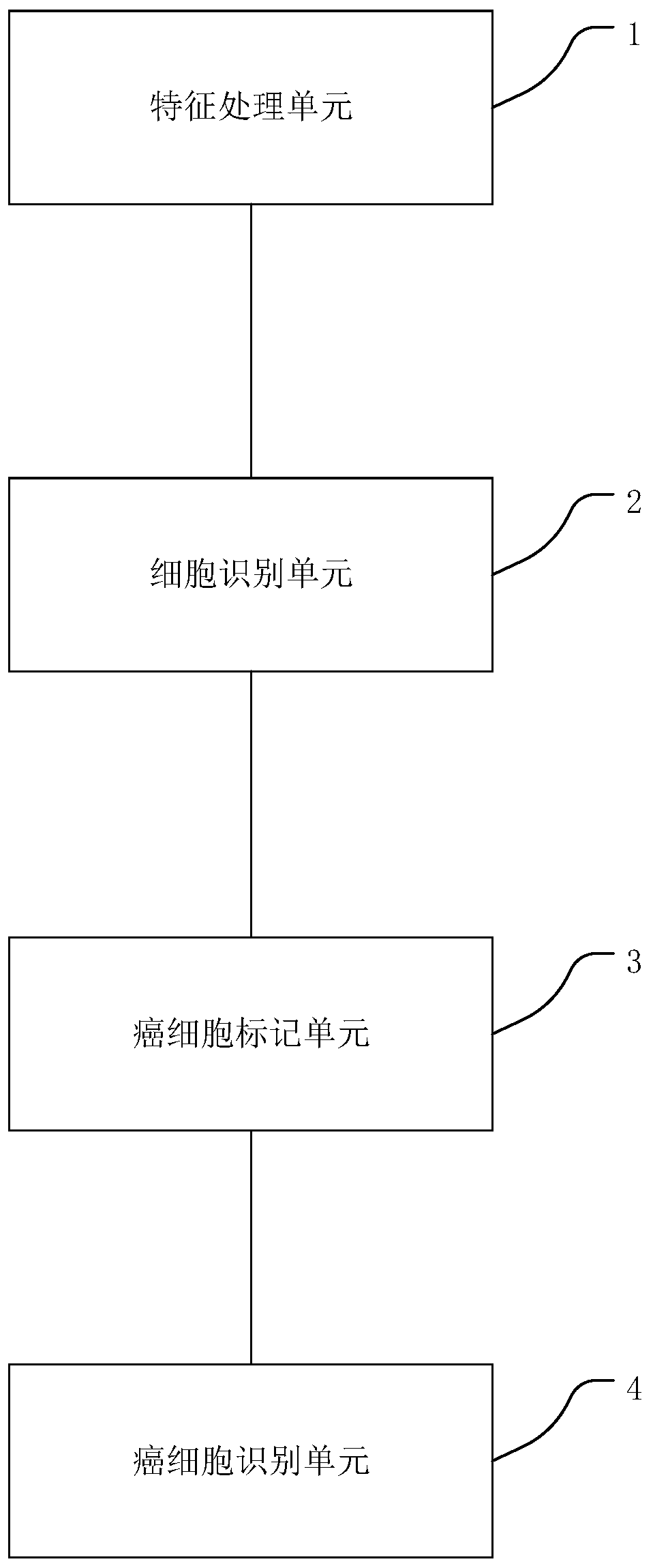
[0074] This embodiment provides as figure 2 A cancer cell identification device shown includes: a feature processing unit 1, configured to process a cell image to be detected based on a residual network to obtain a first feature map; a cell identification unit 2, configured to process the image based on a region proposal network The first feature map obtains a set of elliptical cell candidate frames; the cancer cell labeling unit 3 is configured to process the set of cell candidate frames based on ROIAlign to obtain a second feature map, and process the first feature map through a preset fully connected network. Two feature maps to obtain cancer cell detection frames and cancer cell codes for distinguishing cancer cells; cancer cell identification unit 4, for processing the cancer cell detection frames based on a preset deep learning network to output for marking cancer cell types type information.
[0075] The cancer cell labeling unit 3 is configured to process the second ...
PUM
 Abstract
Description
Claims
Application Information
 - R&D
- Intellectual Property
- Life Sciences
- Materials
- Tech Scout
- Unparalleled Data Quality
- Higher Quality Content
- 60% Fewer Hallucinations
Browse by: Latest US Patents, China's latest patents, Technical Efficacy Thesaurus, Application Domain, Technology Topic, Popular Technical Reports.
© 2025 PatSnap. All rights reserved.Legal|Privacy policy|Modern Slavery Act Transparency Statement|Sitemap|About US| Contact US: help@patsnap.com


