
Electrocoagulation surgical instrument

A surgical instrument and electrocoagulation technology, applied in the field of medical equipment, can solve the problems of frequently inserting and withdrawing electrocoagulation cutters and flushing suction devices into and out of the abdominal cavity, increasing the risk of surgical infection for patients, and low surgical efficiency. The effect of timeliness of treatment, reduction of risk of surgical infection, and convenient operation

Pending Publication Date: 2019-09-24
ZHEJIANG GEYI MEDICAL INSTR
View PDF0 Cites 5 Cited by
  • Summary
  • Abstract
  • Description
  • Claims
  • Application Information

AI Technical Summary

Problems solved by technology

The existing electrocoagulation cutter and flushing aspirator are two independent instruments. When the electrocoagulation operation needs to be performed through the electrocoagulation cutter, electrocoagulation smoke will be continuously generated in the abdominal cavity. When the smoke is condensed to a certain extent, the endoscopic The field of view of the mirror feedback image is affected. At this time, the doctor needs to stop the electrocoagulation, pull the electrocoagulation cutter out of the abdominal cavity, and extend the flushing suction device into the abdominal cavity to suck out the smoke, and then extend the electrocoagulation cutter into the abdominal cavity to continue the electrocoagulation operation , this process needs to frequently insert and withdraw the electrocoagulation cutter and irrigation suction device into and out of the abdominal cavity, which is cumbersome to operate, low in operation efficiency, and increases the risk of surgical infection for patients
For example, in laparoscopic cholecystectomy, the doctor needs to cut the gallbladder quickly and suck the bile out of the body quickly, which cannot be achieved by the existing technology.

Method used


Image

  • Electrocoagulation surgical instrument
  • Electrocoagulation surgical instrument
  • Electrocoagulation surgical instrument

Examples


Embodiment Construction

[0037] In the following, the present invention will be described in detail and completely through specific embodiments in conjunction with the accompanying drawings.

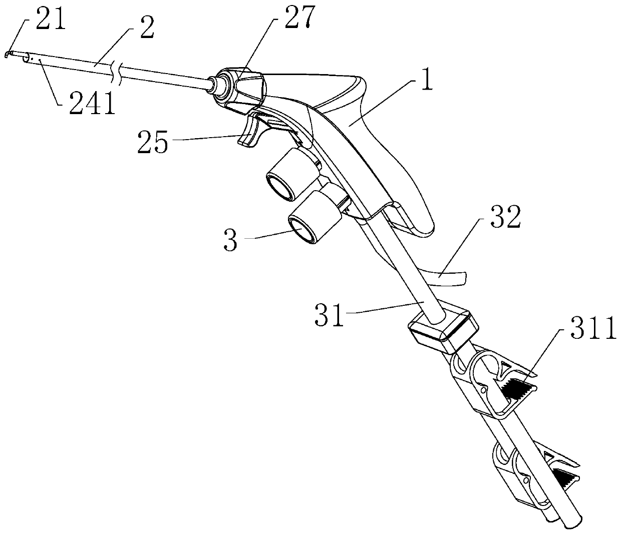
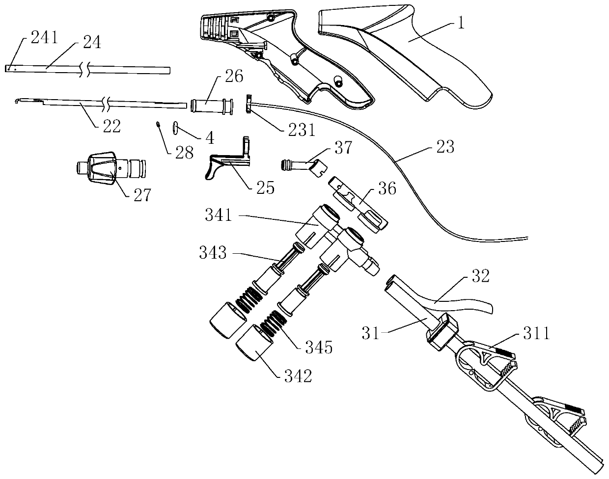
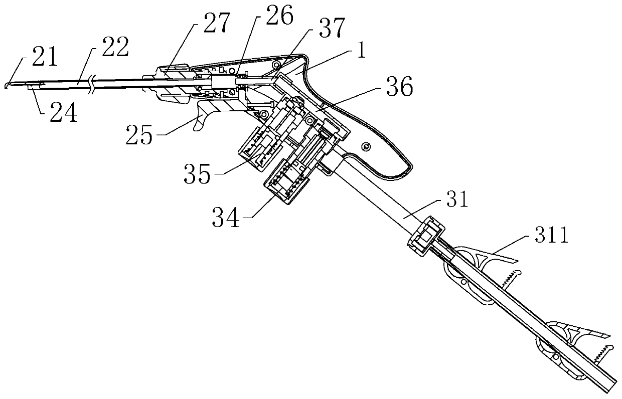
[0038] Please refer to Figure 1-3 As shown, the present invention provides an electrocoagulation surgical instrument, comprising:

[0039] The handle 1 is provided with an installation cavity;

[0040] The electrocoagulation head assembly 2 includes an electrocoagulation head 21, a hollow metal conduit 22 and an electrocoagulation wire 23. The electrocoagulation head 21 is arranged at one end of the metal conduit 22, and the other end of the metal conduit 22 extends into the installation cavity, and the electrocoagulation wire 23 extends into the installation cavity and connects with the metal conduit 22, and the electrocoagulation wire 23 is used to transmit electric signals of electrocoagulation or cutting to the electrocoagulation head 21; and

[0041] The flushing suction assembly 3 includes a flushing tu...


PUM

PropertyMeasurementUnit
Angleaaaaaaaaaa

Abstract

The invention discloses an electrocoagulation surgical instrument. The electrocoagulation surgical instrument comprises a handle, an electrocoagulation head assembly, and a flushing suction assembly; a mounting cavity is formed in the handle; the electrocoagulation head assembly comprises an electrocoagulation head, a metal conduit and an electrocoagulation wire, wherein the electrocoagulation head is arranged at one end of the metal conduit, the other end of the metal conduit extends into the mounting cavity, and the electrocoagulation wire extends into the mounting cavity and is connected to the metal conduit for transmitting electrical signals to the electrocoagulation head; the flushing suction assembly comprises a flushing pipe, a suction pipe, and a pipeline switch, wherein the flushing pipe and the suction pipe extend into the handle and are in communication with the metal conduit, and the pipeline switch is used for controlling the conduction and breaking of the flushing pipe and the suction pipe. The electrocoagulation surgical instrument integrates the functions of electrocoagulation, flushing, and smoke suction, and does not need frequently protruding into and out of the abdominal cavity, so that the risk of surgical infection of patients is reduced; the electrocoagulation surgical instrument can be operated by one hand, achieves rapid conversion of electrocoagulation, flushing, and smoke suction, greatly reduces the time of instrument replacement, is convenient to operate, greatly improves the efficiency of surgery, and improves the timeliness of treatment.

Description

technical field [0001] The invention relates to the technical field of medical instruments, in particular to an electrocoagulation surgical instrument. Background technique [0002] With the development of science and technology, on the premise of ensuring safety and effectiveness, medical devices are developing along the mainstream direction of minimizing surgical trauma, prompting medical devices that realize related operations to pursue more minimally invasive clinical effects. Laparoscopic surgery has been used clinically for decades. Compared with traditional surgery, it has the advantages of less trauma, lower infection rate, and faster postoperative recovery. It has been widely used in urology, gynecology, and abdominal surgery. Laparoscopic surgery is to inject CO into the abdominal cavity after penetrating 2-5 laparoscopic trocars in the patient's abdomen. 2 The gas expands the abdominal cavity, and the endoscope and medical instruments enter the abdominal cavity o...

Claims


Application Information

Patent Timeline
IPC IPC(8): A61B18/12
CPCA61B18/12A61B2217/007A61B2218/008
Inventor 徐欢张迎
Owner ZHEJIANG GEYI MEDICAL INSTR
Learn More