Preparation and Application of Ultrasonic Contrast Agents Derived from Natural Cell Membranes

An ultrasound contrast agent and cell membrane technology, applied in the field of biomedicine, can solve the problems of poor repeatability, cumbersome preparation process, short circulation time of targeted ultrasound contrast agent, etc.

Active Publication Date: 2020-11-27
PEKING UNIV
View PDF2 Cites 0 Cited by
  • Summary
  • Abstract
  • Description
  • Claims
  • Application Information

AI Technical Summary

Problems solved by technology

However, there are still many problems that need to be solved: the coupling technology of ultrasound contrast agents and targeting molecules is immature, the preparation process is cumbersome, and the repeatability is poor; the sensitivity of binding between target molecules and receptors is low, and the efficiency of targeting binding needs to be improved ;Targeted ultrasound contrast agents have a short circulation time in the body and are easily cleared by the immune system, etc.
[0005] In the field similar to the development of ultrasound contrast agents, such as the field of nanomaterials, they also face the problems of poor safety and insufficient functionality

Method used


Image

  • Preparation and Application of Ultrasonic Contrast Agents Derived from Natural Cell Membranes
  • Preparation and Application of Ultrasonic Contrast Agents Derived from Natural Cell Membranes
  • Preparation and Application of Ultrasonic Contrast Agents Derived from Natural Cell Membranes

Examples


Embodiment 1

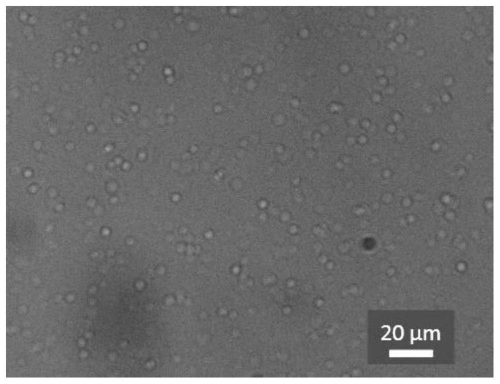
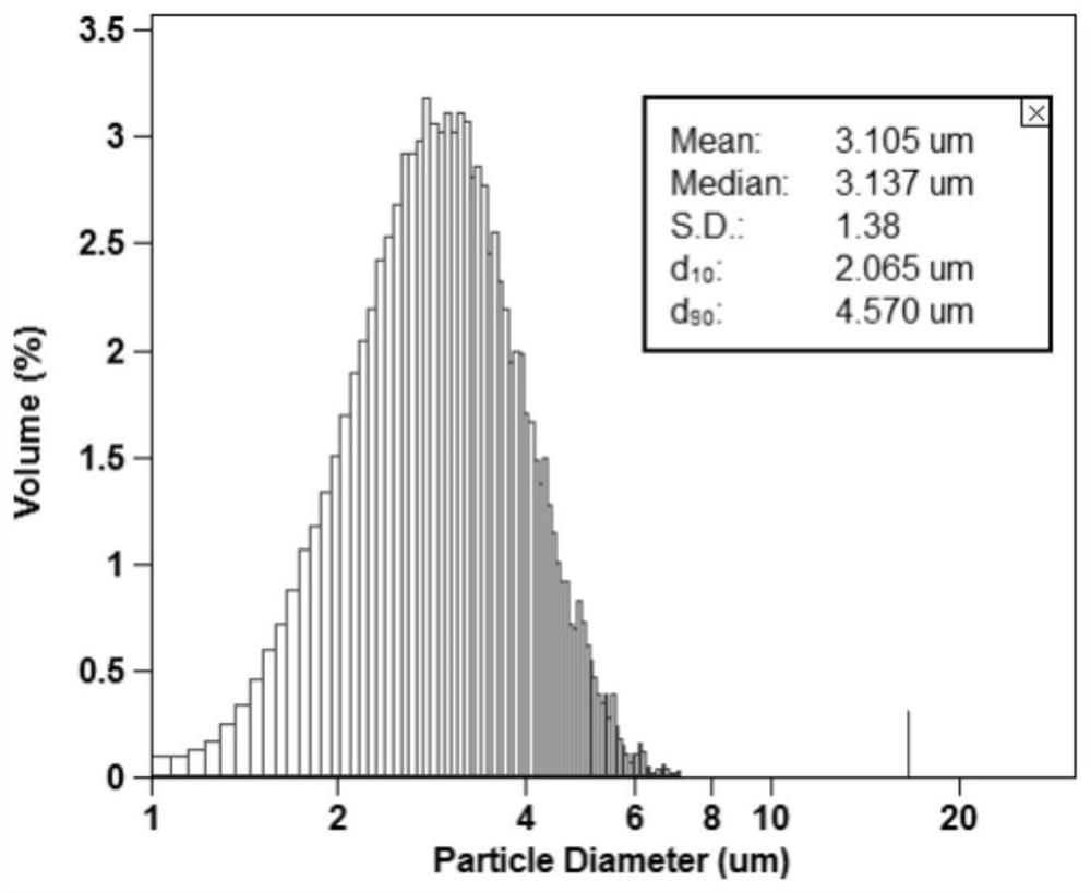
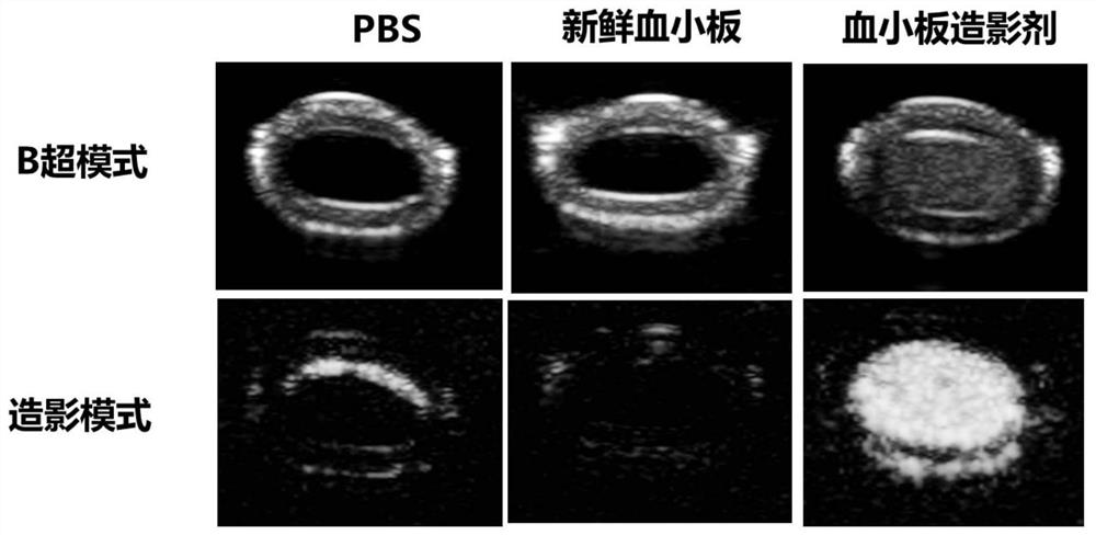
[0024] Preparation of platelet ultrasound contrast agent: extract a certain amount of peripheral anticoagulated venous blood into a sterile centrifuge tube, record the initial volume (V 0 mL). Platelet-rich plasma was prepared by two-step centrifugation. For the first time, a centrifugal force of 200×g was used for 10 minutes. Carefully draw the upper layer of plasma to another centrifuge tube and discard the lower layer of red blood cells. The second centrifugation adopts a centrifugal force of 1390×g for 15 minutes. The platelet plasma volume needs to be preserved according to the calculation of retaining 1 / 10 of the initial volume. Discard the excess plasma in the upper layer, and blow the platelet sediment in the lower layer to obtain high-concentration platelet plasma.

[0025] The freshly separated platelet plasma was centrifuged at 12000×g for one second to separate platelets and upper plasma. Record the partial volume of plasma (V 1 mL). Then the lower platelet ...

Embodiment 2

[0027] Preparation of erythrocyte ultrasound contrast agent: draw a certain amount of peripheral anticoagulated venous blood into a sterile centrifuge tube, centrifuge the blood at 2500r / min for 5min, discard the plasma and buffy coat, and wash the erythrocytes with isotonic pH=7.4 phosphate The buffer solution was centrifuged at 2500r / min for 5min, washed repeatedly for 3 times, and the supernatant was discarded to obtain red blood cells; the obtained washed red blood cells were mixed with 800mM trehalose solution at a ratio of 1:1.5, and the seaweed The sugar is loaded into the cells, so that the concentration of trehalose in the cells reaches about 60mM, and the erythrocyte concentrate of trehalose is obtained. Red blood cell concentrate and lyophilization buffer (15% PVP + 4% trehalose + 3% glycerol + 5% BSA + 0.05% HSI) were mixed according to the volume ratio of 1:4, then freeze-dried after aliquoting, and carried out with nitrogen Backfill, seal and store to obtain the ...

Embodiment 3

[0029] Preparation of ultrasound contrast agent for prostate cancer cells (PC-3): collect mammalian sample cells suspended in albumin in 1% phosphate buffer at 4° C., and centrifuge the cell suspension to obtain cell pellets. Cells were resuspended in trehalose isotonic solution and co-incubated for 1 h. Add the cell suspension to the container, cool to 4°C and shake the container to spread the cells evenly and freeze at about -70°C for 1 h. Transfer the container to a lyophilizer for lyophilization and inert gas SF 6 Backfilling is carried out, sealed and preserved to obtain the freeze-dried powder of prostate cancer cell ultrasound contrast agent.


PUM


Abstract

The invention relates to a natural cell membrane derived novel ultrasonic contrast agent, in particular to a preparation method of the ultrasonic contrast agent and an application of the ultrasonic contrast agent to the respects of diagnosis and treatment of various diseases of cardiovascular diseases, cancer and the like. The ultrasonic contrast agent is characterized in that membrane components are completely from natural cell membranes, then gas or liquid is wrapped or loaded in the membrane components, and the novel contrast agent can approximately maintain structural integrity and function activity of the natural cell membranes. The novel contrast agent can maintain original function activity of the cell membranes, so that the novel contrast agent has the capacity of efficiently avoiding immune elimination in vivo, and blood long circulation capacity, and has favorable active target properties.

Description

technical field [0001] The invention belongs to the field of biomedicine, and in particular relates to the preparation of a novel ultrasound contrast agent derived from natural cell membranes and its application in the diagnosis and treatment of various diseases such as cardiovascular disease and cancer. Background technique [0002] Non-invasive ultrasound imaging has been widely used in the diagnosis of various diseases such as cancer and cardiovascular diseases because of its incomparable advantages of convenience, low cost and real-time imaging. Compared with other imaging methods, ultrasonography is painless, radioactive, and does not require special environmental conditions. It provides accurate, rich, and intuitive information, and is widely loved by patients and doctors. Due to the economical and practicality of ultrasonography, the popularity of ultrasonic diagnostic equipment in my country is much higher than that of expensive imaging techniques such as CT and magn...

Claims


Application Information

Patent Timeline
Patent Type & Authority Patents(China)
IPC IPC(8): A61K49/22
CPCA61K49/223
Inventor 戴志飞高闯徐云雪周一鸣
Owner PEKING UNIV
Why Eureka
Try Eureka