Method, device and equipment for processing image

An image and medical equipment technology, applied in the field of image processing, which can solve the problems of patients and doctors, such as physical damage and large damage to the human body.

Active Publication Date: 2019-10-08
NEUSOFT MEDICAL SYST CO LTD +1
View PDF16 Cites 2 Cited by
  • Summary
  • Abstract
  • Description
  • Claims
  • Application Information

AI Technical Summary

Problems solved by technology

[0004] The vascular machine uses X-rays (X-ray) for scanning and exposure. Since X-rays are more harmful to the human body, multiple scanning and exposure processes will cause greater physical damage to patients and doctors.

Method used


Image

  • Method, device and equipment for processing image
  • Method, device and equipment for processing image
  • Method, device and equipment for processing image

Examples


Embodiment Construction

[0104] Reference will now be made in detail to the exemplary embodiments, examples of which are illustrated in the accompanying drawings. When the following description refers to the accompanying drawings, the same numerals in different drawings refer to the same or similar elements unless otherwise indicated. The implementations described in the following exemplary examples do not represent all implementations consistent with the present invention. Rather, they are merely examples of apparatuses and methods consistent with aspects of the invention as recited in the appended claims.

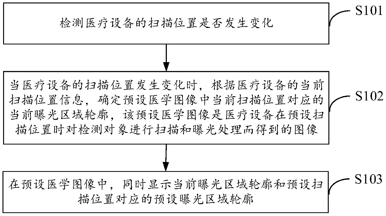
[0105] figure 1 It is a flowchart of a method for processing an image according to an exemplary embodiment. The method is applied to medical equipment, and the medical equipment obtains a medical image by scanning and exposing a detection object. The method includes:

[0106] In step S101, it is detected whether the scanning position of the medical device changes.

[0107] The medical equipmen...


PUM


Abstract

The invention relates to a method, device and equipment for processing an image. The method for processing the image is applied to medical equipment. The method for processing the image comprises thesteps that whether a scanning position of the medical equipment is changed or not is detected, when the scanning position of the medical equipment is changed, according to current scanning position information of the medical equipment, a current exposure area contour corresponding to the current scanning position in the preset medical image is determined, and the preset medical image is an image obtained by performing scanning and exposure treatment on a detection object when the medical equipment is at the preset scanning position; and in the preset medical image, the current exposure area contour and a preset exposure area contour corresponding to the preset scanning position are simultaneously displayed, so that a doctor quickly locates the lesion by viewing the current exposure area contour in the preset medical image and the preset exposure area contour, the number of scanning and exposure treatment is reduced, the physical damage to the doctor and patient caused by the radiationused by the medical equipment is reduced.

Description

technical field [0001] The present invention relates to the technical field of medical image processing, in particular to a method, device and equipment for image processing. Background technique [0002] When using medical equipment to perform medical image detection on patients, for example, when using a vascular machine to perform medical image detection on patients, the patient needs to lie on the test bed of the vascular machine, and the doctor controls the movement of the vascular machine frame through the console. The body part is scanned and exposed to obtain a medical image of the body part to be detected. [0003] In the related technology, after the patient lies on the test bed, the doctor controls the frame of the blood vessel machine to move to the vicinity of the body part of the patient to be tested according to the operating experience, and then performs multiple scans and exposure processing on the patient, and obtains multiple medical images obtained by vie...

Claims


Application Information

Patent Timeline
Patent Type & Authority Applications(China)
IPC IPC(8): A61B6/00
CPCA61B6/00A61B6/4476A61B6/504A61B6/52A61B6/54
Inventor 郝华明孙艳虎王静刘建勋
Owner NEUSOFT MEDICAL SYST CO LTD
Why Eureka
Try Eureka