Intraoperative tissue cherenkov fluorescence imaging system and image processing method thereof

A technology of fluorescence imaging and image processing, which is applied in the fields of analysis using fluorescence emission, medical science, sensors, etc. It can solve the problems of long analysis time and small area of ​​frozen pathological sections, and achieve high signal-to-background ratio and efficient tissue margins The effect of fast inspection and imaging speed

Inactive Publication Date: 2019-10-15
INST OF AUTOMATION CHINESE ACAD OF SCI
View PDF5 Cites 0 Cited by
  • Summary
  • Abstract
  • Description
  • Claims
  • Application Information

AI Technical Summary

Problems solved by technology

[0006] In order to solve the above-mentioned problems in the prior art, that is, the problem of small sampling area and long analysis time of intraoperative frozen pathological sections, t

Method used


Image

  • Intraoperative tissue cherenkov fluorescence imaging system and image processing method thereof
  • Intraoperative tissue cherenkov fluorescence imaging system and image processing method thereof
  • Intraoperative tissue cherenkov fluorescence imaging system and image processing method thereof

Examples


Embodiment Construction

[0042] The application will be further described in detail below in conjunction with the accompanying drawings and embodiments. It should be understood that the specific embodiments described here are only used to explain related inventions, not to limit the invention. It should also be noted that, for the convenience of description, only the parts related to the related invention are shown in the drawings.

[0043] It should be noted that, in the case of no conflict, the embodiments in the present application and the features in the embodiments can be combined with each other. The present application will be described in detail below with reference to the accompanying drawings and embodiments.

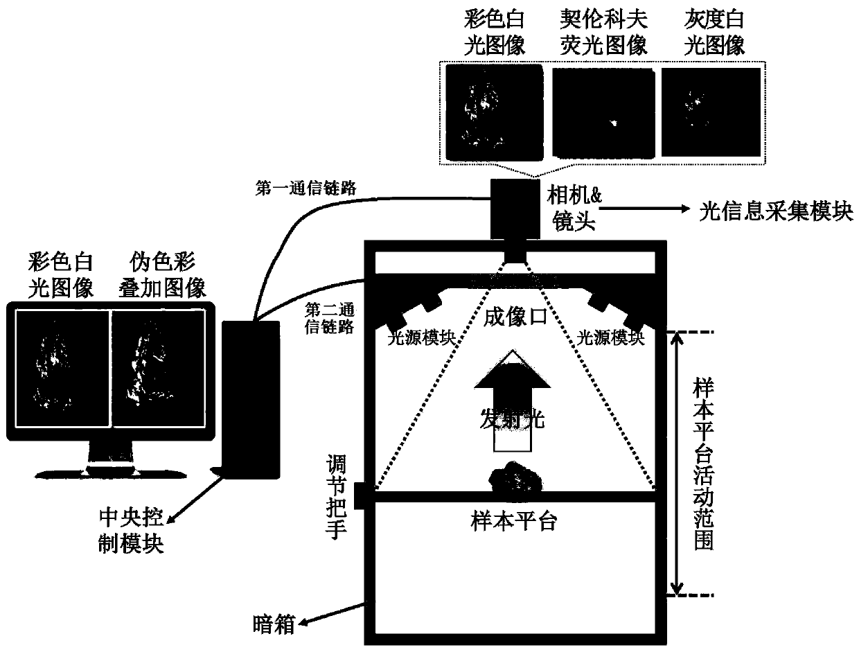
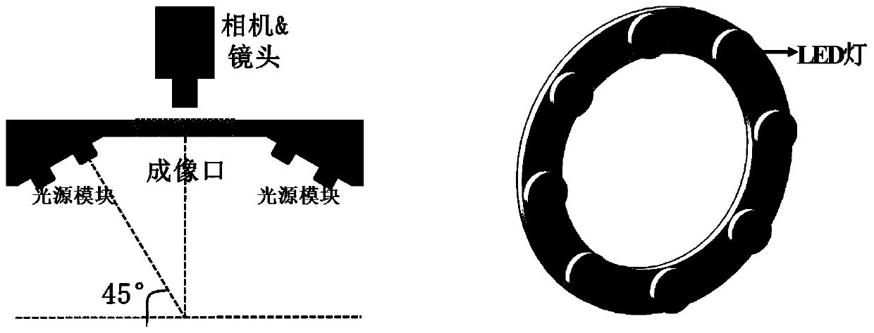
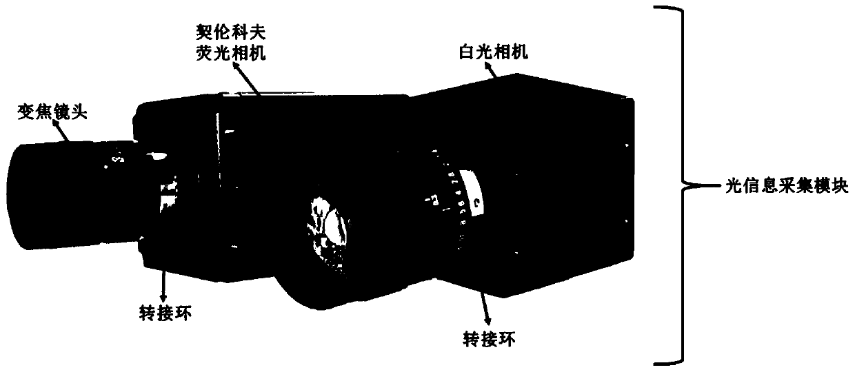
[0044] An intraoperative tissue Cherenkov fluorescence imaging system of the present invention, the fluorescence imaging system includes a light source module, an information collection module, and a central control module;

[0045] The light source module includes a white light emi...


PUM


Abstract

The invention belongs to the field of optical molecule imaging, in particular to an intraoperative tissue cherenkov fluorescence imaging system and an image processing method thereof. The problems that the material collection area of intraoperative frozen pathologic slices is small and the analysis time is long are solved. The system disclosed by the invention comprises a light source module, an information acquisition module, and a central control module, wherein the light source module is used for emitting white light irradiated intraoperative tissues; the information acquisition module is used for acquiring colored white light images, gray-scale white light images and cherenkov fluorescence images of the intraoperative tissues; the central control module is used for controlling the light source module to lighten and controlling the information acquisition module to acquire the images and receive the state information of the light source module and image information acquired by the information acquisition module; and the intraoperative tissue images are generated through a built-in image processing algorithm. According to the imaging system disclosed by the invention, spontaneoussignals are produced during imaging, a signal-to-background ratio is high, and an imaging process is high in specificity, high in sensitivity, high in resolution ratio of a shallow layer and high inimaging speed; and moreover, surgical margin testing of the intraoperative tissues can be quickly and effectively realized.

Description

technical field [0001] The invention belongs to the field of optical molecular imaging, and in particular relates to an intraoperative tissue Cerenkov fluorescence imaging system and an image processing method thereof. Background technique [0002] According to China's cancer statistics, the incidence and mortality of colorectal cancer rank among the top, seriously threatening the national health. In my country, the proportion of patients with low rectal cancer is over 65%, and the conventional treatment method is mainly surgical resection. In order to improve the postoperative survival period and quality of life of patients, it is necessary to detect the boundaries of the resected tissue during the operation to ensure that the tumor tissue is completely resected while retaining the normal rectal tissue of the patient as much as possible. In current clinical practice, intraoperative frozen pathology is often used to detect the boundary of the resected tumor tissue. If the b...

Claims


Application Information

Patent Timeline
IPC IPC(8): A61B5/00
CPCA61B5/0071
Inventor 田捷胡振华张泽宇史小静曹财广
Owner INST OF AUTOMATION CHINESE ACAD OF SCI
Why Eureka
Try Eureka