
Method for automatically extracting central paths of cephalic and cervical blood vessels in CTA image

An automatic extraction, center path technology, applied in cardiac catheterization, instruments for radiological diagnosis, medical science, etc., can solve the problems of vascular disease analysis, loss or fracture of vascular structure, easy displacement of patients, etc., and achieve fast operation speed. , the center line is accurate, and the effect of meeting real-time requirements

Active Publication Date: 2019-10-18
SAINUO WEISHENG SCI & TECH BEIJING
View PDF6 Cites 6 Cited by
  • Summary
  • Abstract
  • Description
  • Claims
  • Application Information

AI Technical Summary

Problems solved by technology

The disadvantage of this method is: since the subtraction operation requires two scans (CT and CTA scans) of the patient, the patient is prone to displacement during the interval between the two scans, which will cause the loss of the vascular structure after subtraction or rupture, which will affect the follow-up analysis of vascular lesions
[0004] Using the Hessian-based blood vessel enhancement method to obtain the central path of blood vessels, although the central path of blood vessels can be directly extracted in the CTA image, it is more complicated for the shape and surrounding tissue For example, the cranial entry of the vertebrae and the internal carotid artery, the effect is usually not very satisfactory, and it is also necessary for the doctor to manually define the starting point and the ending point of the blood vessel, which brings a certain burden to the doctor's analysis work

Method used


Image

  • Method for automatically extracting central paths of cephalic and cervical blood vessels in CTA image
  • Method for automatically extracting central paths of cephalic and cervical blood vessels in CTA image
  • Method for automatically extracting central paths of cephalic and cervical blood vessels in CTA image

Examples


Embodiment Construction

[0020] The present invention will be described in detail below in conjunction with the accompanying drawings and embodiments. It should be noted that various modifications can be made to the embodiments disclosed herein, therefore, the embodiments disclosed in the specification should not be regarded as limitations on the present invention, but only as examples of embodiments, and its purpose is to make the present invention The features of the invention are self-evident.

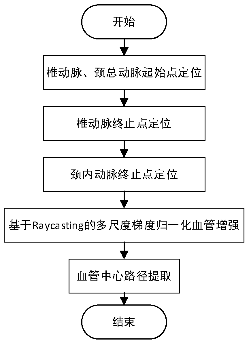
[0021] Such as figure 1 As shown, the method for automatically extracting the central path of the head and neck vessels (vertebral artery and carotid artery) in the CTA image provided by the present invention is applicable to the CTA image data scanned from the aortic arch to the top of the skull (hereinafter described as CTA image data meeting the requirements). Method provided by the invention mainly comprises three key steps:

[0022] S1: According to the shape, grayscale and position characteristics o...


PUM


Abstract

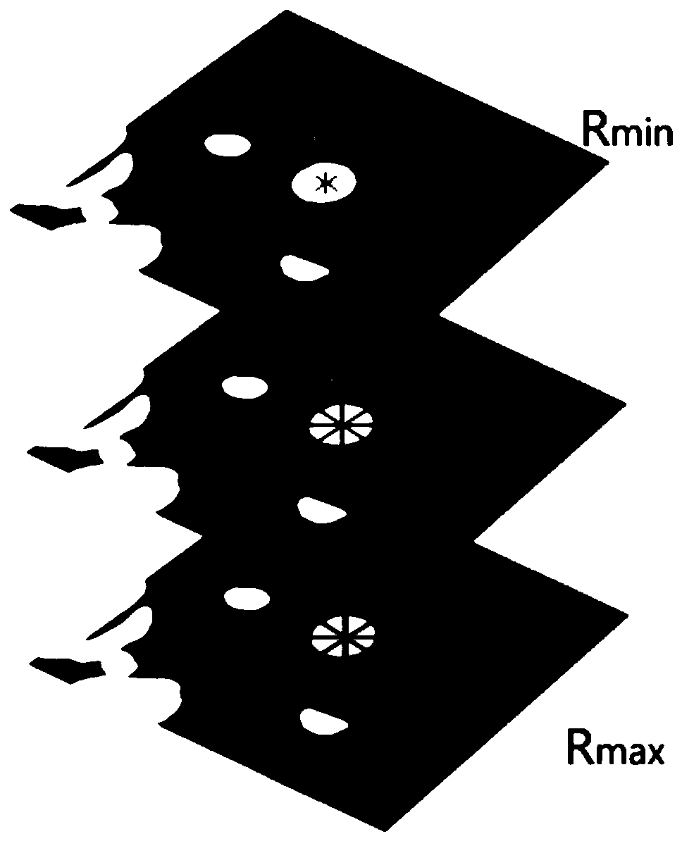
The invention provides a method for automatically extracting the central paths of cephalic and cervical blood vessels in a CTA image. The method is applicable to CTA image data scanned from the aorticarch to the calva, and includes the following steps that S1, according to the shape, gray and position features of the cross sections of the blood vessels in the CTA image, the starting and ending points of the carotid artery and the vertebral artery are automatically localized; S2, between the localized starting and ending points of the blood vessels in step S1, Raycasting-based multi-scale gradient normalization blood vessel filtering is conducted on areas meeting the blood vessel gray features to construct a blood vessel filtering weight map; S3, in the blood vessel filtering weight map constructed in step S2, extraction of the central paths of the carotid artery and the vertebral artery is completed by adopting a Dijkstra optimal path extraction algorithm. The method has the advantages that automatic localization of the blood vessels is realized without manual operation of a user; the extracted center lines are accurate, and the user does not need to perform later adjustment; themethod runs fast and meets the requirement for real-time performance.

Description

technical field [0001] The invention relates to a method for automatically extracting the central path of head and neck vessels in CTA images. The invention belongs to the technical field of medical image processing. Background technique [0002] Cerebrovascular disease is one of the major diseases that currently threaten human health. In clinical practice, it mainly analyzes the images after CTA images (ie, non-traumatic vascular imaging examinations) and extracts the central path of blood vessels to obtain the three-dimensional structure information of blood vessels. Doctors judge the degree of vascular disease. However, due to the complex distribution of blood vessels in the human body, blood vessels often pass through the bones, and at the same time, the bones and blood vessels in the images generated after CTA images show bright gray features, which makes it difficult for the extraction of vascular center paths and pathological changes. Analysis poses great difficulti...

Claims


Application Information

Patent Timeline
Patent Type & Authority Applications(China)
IPC IPC(8): A61B6/00A61B6/03
CPCA61B6/504A61B6/501A61B6/5258A61B6/032
Inventor 孙奇
Owner SAINUO WEISHENG SCI & TECH BEIJING
Learn More