Chest X-ray image intelligent diagnosis system and diagnosis method thereof

An intelligent diagnosis and chest technology, applied in the field of medical information, can solve the problem of low reporting efficiency, and achieve the effect of improving diagnosis efficiency, reducing workload, and reducing workload.

Inactive Publication Date: 2019-12-10
北京赛迈特锐医疗科技有限公司
View PDF7 Cites 8 Cited by
  • Summary
  • Abstract
  • Description
  • Claims
  • Application Information

AI Technical Summary

Problems solved by technology

[0004] In view of this, the main purpose of the present invention is to provide a chest X-ray image intelligent diagnosis system and its diagnosis method, which can solve the problem in the prior art that doctors have to diagnose each image one by one due to the lack of a complete workflow. Inefficiency of doctors diagnosing and writing reports

Method used


Image

  • Chest X-ray image intelligent diagnosis system and diagnosis method thereof
  • Chest X-ray image intelligent diagnosis system and diagnosis method thereof
  • Chest X-ray image intelligent diagnosis system and diagnosis method thereof

Examples


Embodiment 1

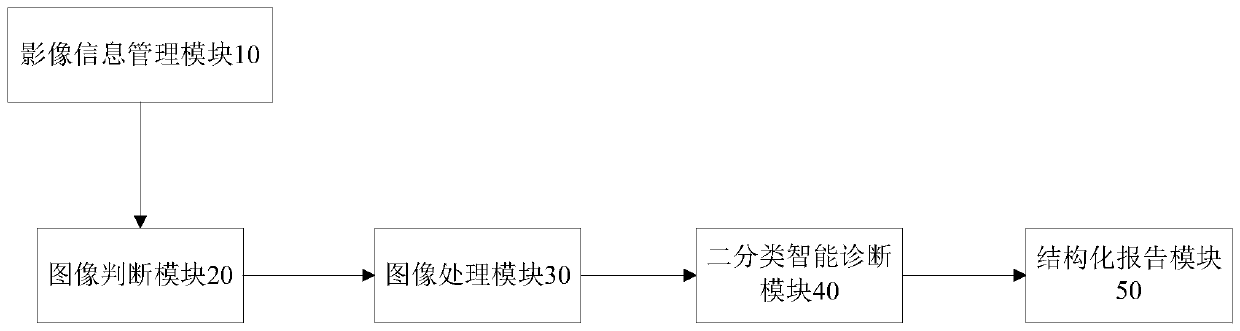
[0033] figure 1 It shows a schematic structural diagram of a chest X-ray image intelligent diagnosis system according to Embodiment 1 of the present invention;figure 1 As shown, the system includes: an image information management module 10, an image judgment module 20, an image processing module 30, a binary classification intelligent diagnosis module 40 and a structured report module 50, wherein,

[0034] The image information management module 10 is connected to the image judging module, and is used to transmit the patient's medical digital imaging and communication DICOM images to the image judging module 20 through the DICOM protocol when the patient has finished taking the inspection item of chest radiograph;

[0035] Among them, the image information management module is RIS (Radiology Information System) system; the types of patients are outpatients, physical examination patients, and preoperative routine examinations for inpatients.

[0036] The image judging module 2...

Embodiment 2

[0046] Figure 4 A schematic structural diagram of a chest X-ray image intelligent diagnosis system according to Embodiment 2 of the present invention is shown, as Figure 4 As shown, the image judging module 20 also includes a feedback unit 202, which is connected to the structured report module 50, and is used to compare the patients corresponding to the non-frontal chest images in the DICOM images and the frontal chest images that do not meet the preset conditions. The unique ID is sent to the structured reporting module for further diagnosis by the doctor.

[0047] For example, the lateral chest radiograph in the received DICOM image is a chest radiograph that does not meet the requirements, or the image quality of the front chest radiograph image is unqualified due to some reasons, which is also a chest radiograph that does not meet the requirements. The feedback unit sends the patient's unique ID corresponding to the chest radiographs that do not meet the requirements t...

Embodiment 3

[0050] Figure 5 It shows a schematic structural diagram of a chest X-ray image intelligent diagnosis system according to Embodiment 3 of the present invention; Figure 5 As shown, the binary classification intelligent diagnosis module 40 includes a feature extraction unit 402 and a calculation unit 404, wherein,

[0051] The feature extraction unit 402 is connected with the calculation unit 404, and is used to extract the feature data of the qualified chest film after processing, and send the feature data to the calculation unit 404;

[0052] The calculation unit 404 is connected with the feature extraction unit 402, and is used to combine the feature data with the fully connected layer trained by a plurality of normal chest films and abnormal chest films to perform combined calculations of the AND or non-relationship of features, and calculate the processed qualified chest When the normal probability is greater than the first preset threshold, the normal data is output; whe...


PUM


Abstract

The invention provides a chest X-ray image intelligent diagnosis system which comprises: an image information management module which transmits a chest radiography examination image of a patient to animage determining module; an image judgment module which is used for identifying a normal position chest radiography image in the image, analyzing the quality of the normal position chest radiographyimage, sending the normal position chest radiography image meeting a preset condition to the image processing module, and defining the normal position chest radiography image as a qualified chest radiography; an image processing module which performs unified processing on the qualified chest radiography; a binary classification intelligent diagnosis module which is used for carrying out normal orabnormal diagnosis on the processed qualified chest radiography; a structured report module which is used for activating a discovering-free related control in a structured report interface when receiving normal data and inactivating the non-discovery related control ; and when abnormal data is received. The invention further discloses a chest X-ray image intelligent diagnosis method. According tothe invention, the chest radiography is intelligently diagnosed into a normal type and an abnormal type through a binary classification method, and the result is sent to the structured report, so that the diagnosis efficiency of doctors is improved.

Description

technical field [0001] The present invention relates to the field of medical information, more specifically, to a chest X-ray image intelligent diagnosis system and a diagnosis method thereof. Background technique [0002] Chest X-ray clinical examination is referred to as chest X-ray, which is widely used clinically. The frontal chest radiograph can show the shape, position and outline of the lungs, mediastinum, diaphragm, chest wall bone and soft tissue, etc. It can observe lung lesions and the outline of the heart and great vessels, and can be used for the diagnosis of diseases mainly related to the respiratory system. CNN can be used for research on intelligent diagnosis of chest X-ray (Chest X Ray, CXR), but there is no mature product. The most important work in the existing research is the multi-class diagnosis of chest X-ray, which some companies use for the automatic detection of diseases such as tuberculosis. [0003] The existing technology does not pay attention...

Claims


Application Information

Patent Timeline
Patent Type & Authority Applications(China)
IPC IPC(8): G06T7/00G16H30/20G16H15/00A61B6/00
CPCA61B6/52G06T7/0012G06T2207/10081G06T2207/20081G06T2207/20084G06T2207/30061G16H15/00G16H30/20
Inventor 岳新王霄英
Owner 北京赛迈特锐医疗科技有限公司
Why Eureka
Try Eureka