Living body sampling device for digestive system department

A sampling device, gastroenterology technology, applied in medical science, extraction and delivery system, inoculation and ovulation diagnosis, etc., can solve the problems of obstructed observation, troublesome medical personnel, stomach or intestinal irritation, etc., and achieve easy observation and sampling, structure The effect of simple and accurate diagnosis

Active Publication Date: 2019-12-17
重庆市人民医院
View PDF24 Cites 7 Cited by
  • Summary
  • Abstract
  • Description
  • Claims
  • Application Information

AI Technical Summary

Problems solved by technology

[0003] When the existing living body sampling device enters the human body, because the temperature is lower than the body temperature, it will bring greater stimulation to the stomach or intestinal tract, causing people's discomfort, and at the same time, there may be fluid accumulation in the patient's diseased tissue Too many mixtures will hinder the observation, affect the accurate extraction of diseased tissues, and bring great troubles to medical personnel

Method used


Image

  • Living body sampling device for digestive system department
  • Living body sampling device for digestive system department
  • Living body sampling device for digestive system department

Examples


Embodiment 1

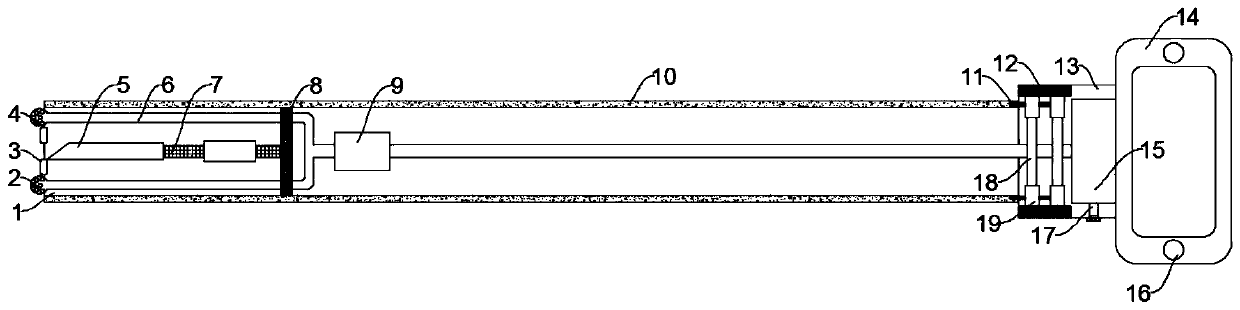
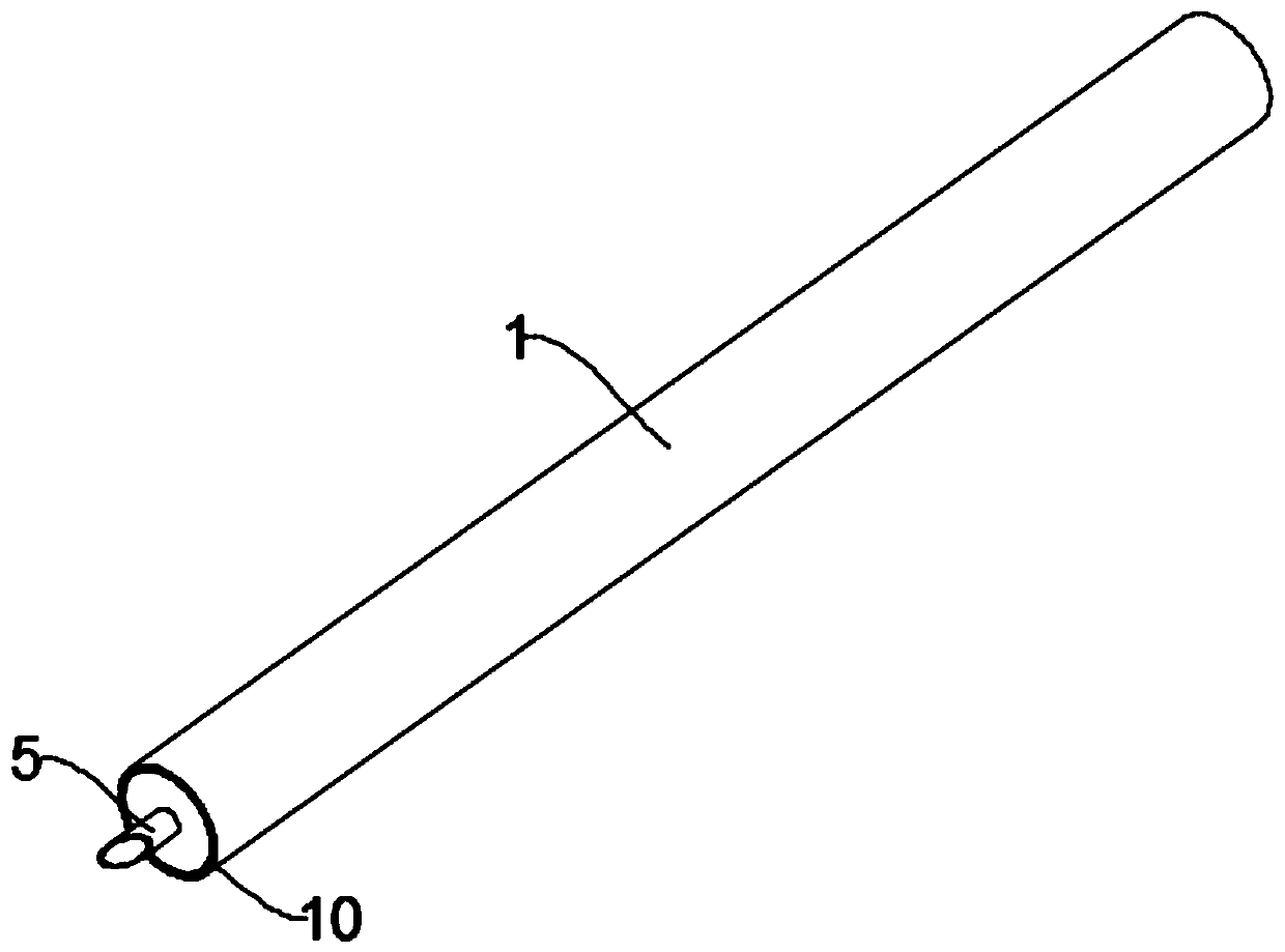
[0024] see figure 1 and figure 2 , in an embodiment of the present invention, a living body sampling device for gastroenterology, comprising a cannula 1, an instrument compartment 13 and a handle 14, the outer wall of the cannula 1 is covered with an antibacterial coating, and the side wall of the cannula 1 is a sandwich structure, The interlayer structure of the side wall of the intubation tube 1 is filled with heat-conducting silica gel 10, the instrument compartment 13 is fixedly installed at one end of the intubation tube 1, and a liquid storage box 15 is fixedly installed in the instrument compartment 13, and a liquid outlet is provided on the liquid storage box 15 17, the liquid outlet 17 is provided with a rubber plug, and the instrument compartment 13 is symmetrically provided with a heat insulating block 12, and a heating rod 18 is fixedly installed between the two heat insulating blocks 12, and a heat conduction ring 19 is arranged on the heating rod 18. The heat c...

Embodiment 2

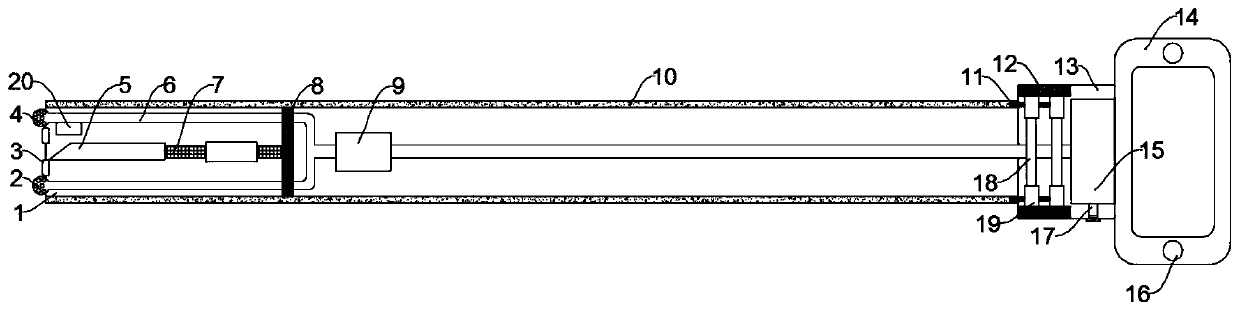
[0026] see image 3 The difference between the embodiment of the present invention and embodiment 1 is that, further, in order to allow medical personnel to understand the pathological condition of the patient's diseased tissue more clearly, a pH detector 20 is fixedly installed in the cannula 1, and the pH detector The detection probe of the meter 20 extends to the outside of the intubation tube 1, and the handle 14 is provided with a display device for displaying the pH value. The pH detector 20 is electrically connected to the display device, and the pH detector 20 can feed back the pH of the diseased tissue of the patient to the display. The device is used as a reference for medical personnel, so as to diagnose the disease more accurately, which is simple and practical.

[0027] The working principle of the present invention is: when the present invention is in use, start the heating rod 18 first, the heat generated by the heating rod 18 is transmitted to the heat conducti...


PUM


Abstract

The invention discloses a living body sampling device for the digestive system department. The device comprises a cannula, an instrument bin and a handle, the side wall of the cannula is of a sandwichstructure, the interlayer structure of the side wall of the cannula is filled with heat-conducting silica gel, the instrument bin is fixedly mounted at one end of the cannula, a liquid storage box isfixedly mounted in the instrument bin, heat insulation blocks are symmetrically arranged in the instrument bin, a heating rod is fixedly installed between the two heat insulation blocks, a heat conduction ring is arranged on the heating rod, a heat conduction wire is connected to the heat conduction ring, a liquid guide pipe is arranged in the cannula, a suction pump is arranged on the liquid guide pipe, a fixed block is fixedly installed in the cannula, a telescopic rod is fixedly installed on the fixed block, and a sampling needle is fixedly connected to one end, away from the fixed block,of the telescopic rod, the handle is fixedly installed at the end, away from the cannula, of the instrument bin, and the handle is provided with a control button used for controlling the device. The device is adjustable in temperature, more comfortable for a patient to use, and capable of effectively removing hydrops in human tissues, facilitates more accurate observation and sampling for medicalpersonnel, and is simple in structure, convenient and practical.

Description

technical field [0001] The invention relates to the technical field of medical devices, in particular to a living body sampling device for gastroenterology. Background technique [0002] Gastroenterology is a third-level clinical discipline that mainly studies diseases of the esophagus, stomach, small intestine, large intestine, liver, gallbladder, and pancreas. Endoscopic examination is a commonly used clinical examination method because it can visually observe gastrointestinal lesions. In the process of endoscopic examination, doctors often use a living tissue sampling device to first pass through the endoscope to sample living tissue. The device is connected to the patient's body, and a certain amount of diseased tissue is removed from the patient's body by cutting, clamping or puncturing for pathological examination. [0003] When the existing living body sampling device enters the human body, because the temperature is lower than the body temperature, it will bring gre...

Claims


Application Information

Patent Timeline
Patent Type & Authority Applications(China)
IPC IPC(8): A61B10/02G01N33/48A61M1/00
CPCA61M1/0023A61B10/0233G01N33/48
Inventor 蒋昊翔曹毅岳妍
Owner 重庆市人民医院
Why Eureka
Try Eureka