
Method for rapidly separating immunomagnetic beads

An immunomagnetic bead and rapid technology, applied in the field of immunological detection, can solve the problem of rapid chemiluminescence affecting the test speed, etc., and achieve the effect of shortening the time of magnetic separation and cleaning, simplifying the structure and high efficiency.

Inactive Publication Date: 2019-12-17
姜宇腾
View PDF7 Cites 0 Cited by
  • Summary
  • Abstract
  • Description
  • Claims
  • Application Information

AI Technical Summary

Problems solved by technology

Of course, this does not have much impact on the chemiluminescence detection method of the traditional conventional test speed (20-40 minutes), but it seriously affects the test speed of fast chemiluminescence (3-7min).

Method used

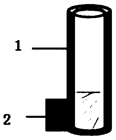
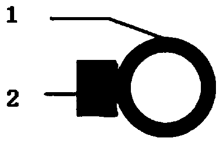
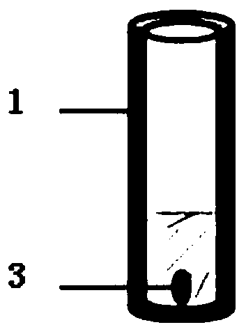

Image

  • Method for rapidly separating immunomagnetic beads
  • Method for rapidly separating immunomagnetic beads
  • Method for rapidly separating immunomagnetic beads

Examples


Embodiment 1

[0044] Acridine ester double monoclonal antibody sandwich chemiluminescence detection, the materials used are: (1) immunomagnetic bead suspension coated with paired monoclonal antibody 1, (2) acridinium ester labeled paired monoclonal antibody 2 solution, (3) Spherical small permanent magnet, (4) exciter solution.

[0045] The details are as follows:

[0046] (1) Add 30 microliters of the serum sample to be tested (or antigen standard 0.01-100.00ng / ml), 100 microliters of immunomagnetic beads suspension coated with antibody 1, and 200 microliters of acridinium ester-labeled monoclonal antibody 2 solution Incubate at 37°C for 3 minutes in a reaction cup;

[0047] (2) Add a small permanent magnet with a diameter of 3 mm into the reaction tube for magnetic separation for 10 seconds, remove the reaction solution, and wash the magnetic particles with cleaning solution for 5 times;

[0048] (3) Add the stimulus solution (first add 100 microliters of 0.1 mol / L HNO 3 +0.1%H 2 o 2...

Embodiment 2

[0051] Alkaline phosphatase competitive chemiluminescence detection, the materials used are: (1) immunomagnetic bead suspension coated with antigen-specific reactive monoclonal antibody, (2) antigen solution labeled with alkaline phosphatase, (3) Spherical small permanent magnet, (4) luminescent substrate solution.

[0052] The details are as follows:

[0053] (1) Add 30 microliters of the serum sample to be tested (or antigen standard 0.01-100.00ng / ml), 100 microliters of antibody-coated immunomagnetic bead suspension, and 200 microliters of alkaline phosphatase-labeled antigen solution to the reaction Incubate at 37°C for 3 minutes;

[0054] (2) Add a small permanent magnet with a diameter of 3 mm into the reaction tube for magnetic separation for 10 seconds, remove the reaction solution, and wash the magnetic particles with cleaning solution for 5 times;

[0055] (3) Add 150 microliters of luminescence substrate solution (AMPPD-enhancer solution) to carry out luminescence...


PUM

PropertyMeasurementUnit
diameteraaaaaaaaaa

Abstract

The invention discloses a method for rapidly separating immunomagnetic beads. The method can be applied to chemiluminescence instruments and nucleic acid extractors. The method is characterized by comprising the steps of: incubating immunomagnetic beads enveloping antibodies / antigens, the antibodies / antigens marked by markers and a to-be-detected object in a reaction cup; after the reaction is finished, a small permanent magnet is added into the reaction cup to adsorb the immunomagnetic beads, so that an antigen-antibody-magnetic bead immune complex is separated from the reaction liquid; and cleaning the antigen-antibody-magnetic bead immune complex with a cleaning liquid, then adding a luminescent substrate / excitation system into the reaction cup to drive a luminescent substance in the reaction cup to release photons, and detecting luminescent intensity by means of photon counting. According to the method, the small permanent magnet is placed in the reaction liquid, so that the smallpermanent magnet is in direct contact with the immunomagnetic beads in the reaction liquid, rapid separation is realized within an extremely short time (3-15s), the separation speed is high, the efficiency is high, the loss rate of the immunomagnetic beads is significantly reduced, the structure of the magnetic separation module in the instrument design process is greatly simplified, and the timefor separating and cleaning the immunomagnetic beads from the reaction liquid is shortened.

Description

technical field [0001] The invention relates to a method for rapidly separating immunomagnetic beads, which changes from the outside of the reaction cup to the inside of the reaction cup with a small permanent magnet, and belongs to the field of immunological detection. Background technique [0002] Immunological detection methods are a series of experimental methods designed by applying immunological theory to detect antigens, antibodies, immune cells and their secreted cytokines. With the interpenetration of disciplines, the scope of immunology has been continuously expanded, and new immunological detection methods have emerged in an endless stream. The scope of application of immunological methods is also expanding, not only becoming an important method for the diagnosis of various clinical diseases, but also providing convenience for the research of many disciplines. [0003] Chemiluminescence is one of the most important subdivisions in the in vitro diagnostic industry...

Claims


Application Information

Patent Timeline
Patent Type & Authority Applications(China)
IPC IPC(8): G01N33/58G01N33/543
CPCG01N33/54326G01N33/58G01N33/581G01N33/582
Inventor 姜宇腾
Owner 姜宇腾
Learn More