
Magnetic resonance imaging scan fixation and anesthesia integrated device for small animals and application method of integrated device

A nuclear magnetic resonance, small animal technology, applied in the direction of animal restraint equipment, applications, veterinary instruments, etc., can solve the problems of small animals suffocation, death, air leakage, etc., to reduce animal preparation time, and the fixation process is simple. Avoid the effects of suffocation

Active Publication Date: 2020-01-03
HARBIN MEDICAL UNIVERSITY
View PDF21 Cites 1 Cited by
  • Summary
  • Abstract
  • Description
  • Claims
  • Application Information

AI Technical Summary

Problems solved by technology

Tooth rods and anesthesia hoods are made of hard plastic. Excessive force during the connection process or long-term hooking of the teeth for experiments will cause animal teeth to be injured; the anesthesia hood also has the risk of wearing away the tissues near the animal's jaw, and even causes the small animal to suffocate to death; There is a large gap between the anesthesia cover and the mouth of the animal, and there is an air leakage phenomenon, which has potential harm to the environment and the operator
In addition, when fixing the torso of small experimental animals or fixing surface coils, adhesive tape is often used to fix the small animals in the animal bed or the experimental transmitting coil. This fixing method not only requires a lot of adhesive tape, but also leaves sticking matter , not easy to clean; especially for MRI with high requirements for breathing control and posture, it cannot be fixed quickly and effectively
[0005]The temperature of the magnetic resonance imaging magnet room is generally controlled at 20 degrees Celsius, and the active body temperature regulation function of small animals in the anesthetized sleep state is reduced. Long-term low temperature experiments are likely to cause body temperature disorders and even Death, and the experiment was not carried out at a normal physiological temperature of about 37 degrees Celsius, and the experimental data obtained are different from the normal body state

Method used


Image

  • Magnetic resonance imaging scan fixation and anesthesia integrated device for small animals and application method of integrated device
  • Magnetic resonance imaging scan fixation and anesthesia integrated device for small animals and application method of integrated device
  • Magnetic resonance imaging scan fixation and anesthesia integrated device for small animals and application method of integrated device

Examples


Embodiment 1

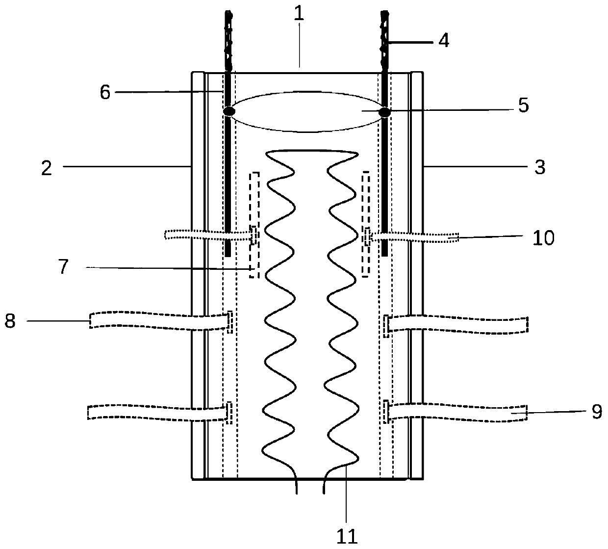
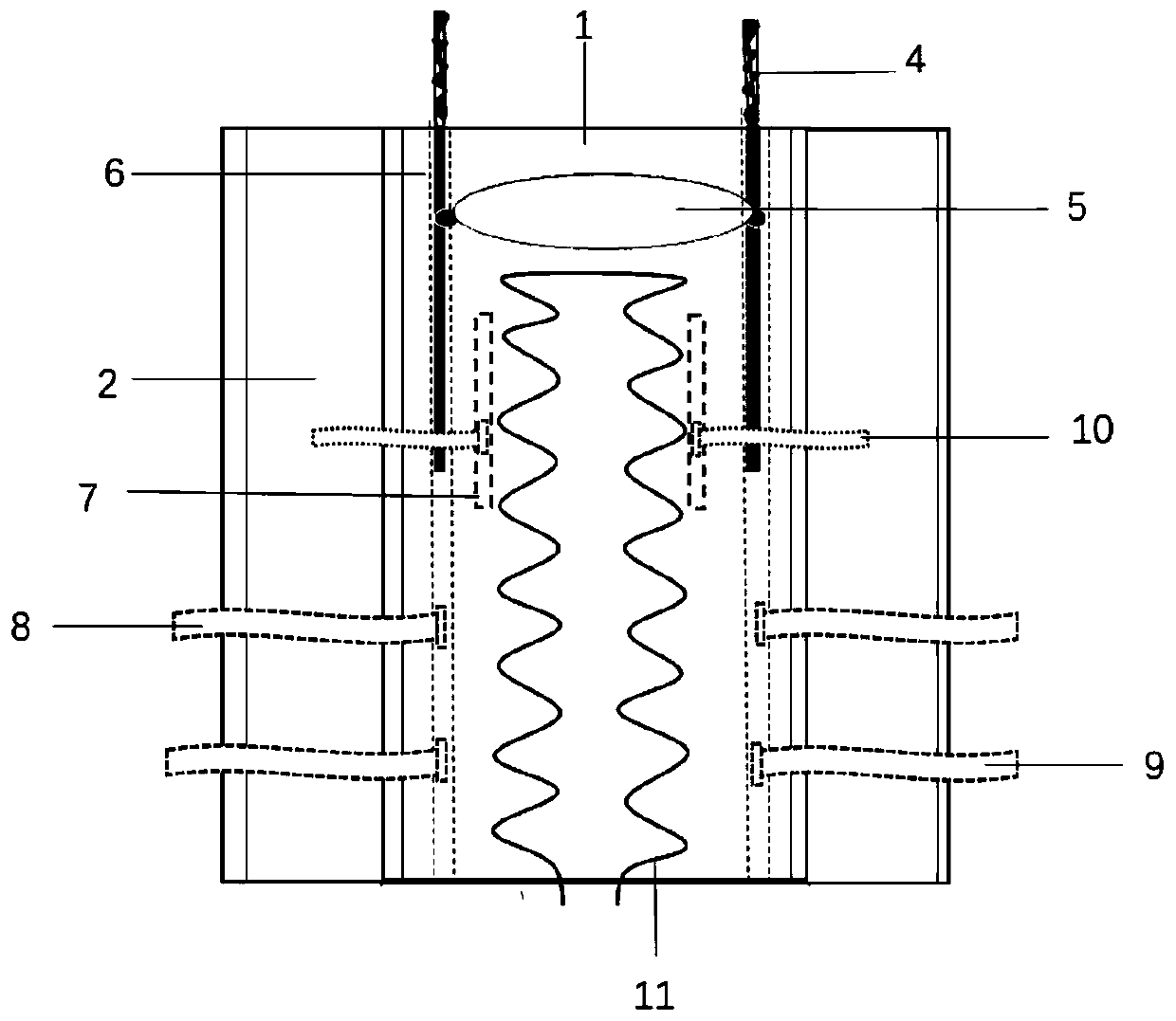
[0038] Such as Figure 1-2 As shown, the small animal MRI scanning fixation and anesthesia integrated device includes an anesthesia module, a head and neck fixation module and a trunk placement fixation module.

[0039] Such as Figure 4 As shown, the anesthesia module includes a cone-shaped inflatable breathing mask, the breathing mask is connected with the outlet pipe of the anesthesia machine through an anesthesia catheter 15, the breathing mask is provided with a first inflation port 16, and the breathing mask is inflated through the inflation port, the breathing mask Elastic rubber bands 14 are fixed on both sides, and the breathing mask is worn on the animal's head after two elastic rubber bands 14 are knotted.

[0040] The head and neck fixation module is detachably connected with the trunk placement fixation module. Such as Figure 5 As shown, the head and neck fixation module includes an adjustable annular inflatable air bag 5, the annular inflatable air bag 5 is s...

Embodiment 2

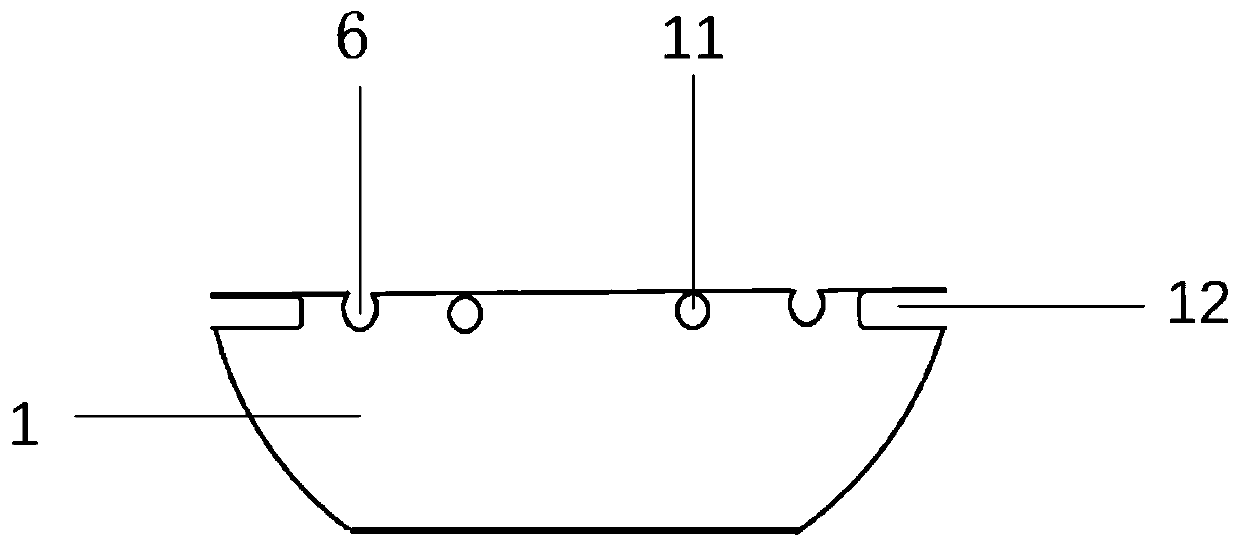
[0046] This embodiment is basically the same as Embodiment 1, the difference is: as Figure 1-3 As shown, both sides of the central body support plate 1 are provided with slots 12 of a hollow structure along the direction in which the animal trunk is placed, and a side trunk support plate 2 is slidably provided in the slot 12, and the side trunk support plate 2 is drawn outwards. The way of pulling changes the width of the fixed module where the torso is placed, which is suitable for fixing animals of different sizes and volumes.

Embodiment 3

[0048] A method for using a small animal nuclear magnetic resonance scanning fixation and anesthesia integrated device includes the following steps:

[0049] (1) Set the water temperature and the flow rate of the water pump in the external heating pool, and connect the two interfaces of the water heating silicone tube 11 with the water outlet and the water return port of the external heating pool;

[0050] (2) quickly place the animal under the initial anesthesia sleep state on the central main body support plate 1, and adjust the side trunk support plate 2 to a suitable position by pulling outward along the slot 12 according to the size of the animal;

[0051] (3) Regulate the inflation rate of the breathing mask by the first inflation port 16, wrap the mouth and nose of the animal with the breathing mask after inflating, after closely fitting, utilize the elastic rubber band 14 that breathing mask carries to tie a knot and wear it on the animal Head, the breathing mask is co...


PUM


Abstract

The invention provides a magnetic resonance imaging scan fixation and anesthesia integrated device for small animals and an application method of the integrated device. The magnetic resonance imagingscan fixation and anesthesia integrated device includes an anesthesia module, a head and neck fixation module and a trunk placement and fixation module, the anesthesia module includes a cone-shaped inflatable breathing mask, and the breathing mask is connected with an external anesthesia tube through an anesthesia catheter; and the head and neck fixation module is detachably connected with the trunk placement and fixation module, and includes an annular inflatable airbag, the annular inflatable airbag is slidably arranged on the trunk placement and fixation module, and a head and neck fixationbelt, an upper waist fixation belt and a lower waist fixation belt are slidably arranged on the trunk placement and fixation module. The magnetic resonance imaging scan fixation and anesthesia integrated device can be adapted to animals with different sizes, put positions of the animals are not restricted, breathing patterns of the animals are conveniently controlled, the body temperature of theanimals can be kept to be stable at the physiological temperature, damage to teeth and tissues near mouths can be avoided, and death from suffocation is effectively avoided.

Description

technical field [0001] The invention relates to the technical field of nuclear magnetic resonance imaging fixation devices, in particular to a small animal nuclear magnetic resonance imaging fixation and anesthesia integrated device and its use method. Background technique [0002] The study of disease model animals can provide reference for the development process of the disease and provide potential solutions for clinical diagnosis and treatment. Magnetic resonance imaging (MRI) has become the preferred imaging detection method for disease diagnosis due to its advantages of non-invasiveness, high resolution, multi-parameter imaging, functional and molecular imaging, and more and more attention has been paid to the study of model animals. [0003] When MRI is performed on animals, gas anesthesia is generally used; animals of different sizes and different scanning parts need to use different radio frequency coils, and different imaging methods and purposes require different ...

Claims


Application Information

Patent Timeline
IPC IPC(8): A61B5/055A61D3/00A61D7/04
CPCA61D3/00A61D7/04A61D2003/003A61D2003/006A61B5/055
Inventor 孙夕林郑利敏王凯杨春升程永娜孙晓红程立欣
Owner HARBIN MEDICAL UNIVERSITY
Learn More